

A new approach to species delimitation in Septoria - CBS - KNAW
A new approach to species delimitation in Septoria - CBS - KNAW
A new approach to species delimitation in Septoria - CBS - KNAW

Create successful ePaper yourself
Turn your PDF publications into a flip-book with our unique Google optimized e-Paper software.
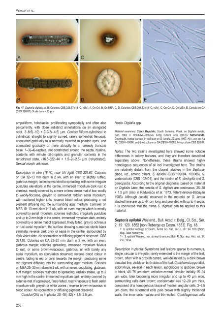
Verkley et al.Fig. 17. Sep<strong>to</strong>ria digitalis. A, B. Colonies <strong>CBS</strong> 328.67 (15 ºC, nUV). A. On OA. B. On MEA. C, D. Colonies <strong>CBS</strong> 391.63 (15 ºC, nUV). C. On OA. D. On MEA. E. Conidia on OA(<strong>CBS</strong> 328.67). Scale bars = 10 µm.ampulliform, holoblastic, proliferat<strong>in</strong>g sympodially and often alsopercurrently, with close <strong>in</strong>dist<strong>in</strong>ct annellations on an elongatedneck, 3–8.5(–10) × 2–3.5(–4.5) µm. Conidia filiform-cyl<strong>in</strong>drical <strong>to</strong>cyl<strong>in</strong>drical, straight <strong>to</strong> slightly curved, rarely somewhat flexuous,attenuated gradually <strong>to</strong> a narrowly rounded <strong>to</strong> po<strong>in</strong>ted apex, andattenuated gradually or more abruptly <strong>to</strong> a narrowly truncatebase, 1–3(–4)-septate, not constricted around the septa, hyal<strong>in</strong>e,contents with m<strong>in</strong>ute oil-droplets and granular contents <strong>in</strong> therehydrated state, (16.5–)22–44 × 1.5–2(–2.5) µm (rehydrated).Sexual morph unknown.Description <strong>in</strong> vitro (18 ºC, near UV light) <strong>CBS</strong> 328.67: Colonieson OA 12–13 mm diam <strong>in</strong> 2 wk, with an even <strong>to</strong> slightly ruffled,glabrous marg<strong>in</strong>; colonies restricted <strong>to</strong> spread<strong>in</strong>g, with some irregularpustulate elevations <strong>in</strong> the centre, immersed mycelium dark rust <strong>to</strong>chestnut, mostly covered by a more or less dense mat of low, woolly<strong>to</strong> woolly-floccose, greyish <strong>to</strong> somewhat reddish aerial mycelium,with scattered higher tufts, reverse blood colour; produc<strong>in</strong>g a redpigment diffus<strong>in</strong>g <strong>in</strong><strong>to</strong> the surround<strong>in</strong>g agar medium. Colonies onMEA 10–13 mm diam <strong>in</strong> 2 wk, with an even marg<strong>in</strong> which is mostlycovered by aerial mycelium; colonies restricted, irregularly pustulateand up <strong>to</strong> 2 mm high <strong>in</strong> the centre, immersed mycelium dark, entirelycovered by a dense mat of appressed, f<strong>in</strong>ely felted, grey <strong>to</strong> ochreousor rust aerial mycelium, the surface show<strong>in</strong>g numerous sterile blackstromata; reverse dark brick or sepia <strong>in</strong> the centre, surrounded bydark violet slate. No sporulation or diffus<strong>in</strong>g pigment observed. <strong>CBS</strong>391.63: Colonies on OA 23–25 mm diam <strong>in</strong> 2 wk, with an even,glabrous marg<strong>in</strong>; colonies spread<strong>in</strong>g, immersed mycelium fulvous<strong>to</strong> rust, or some brown-v<strong>in</strong>aceous, glabrous, or with barely anyaerial mycelium, no sporulation observed; reverse blood colour <strong>in</strong>centre, fad<strong>in</strong>g <strong>to</strong> red or coral <strong>to</strong>wards the marg<strong>in</strong>; produc<strong>in</strong>g somered pigment diffus<strong>in</strong>g <strong>in</strong><strong>to</strong> the surround<strong>in</strong>g agar medium. Colonieson MEA 25–30 mm diam <strong>in</strong> 2 wk, with an even, undulat<strong>in</strong>g, glabrous,buff marg<strong>in</strong>; colonies restricted <strong>to</strong> spread<strong>in</strong>g, radially striate, up <strong>to</strong> 2mm high <strong>in</strong> the centre, immersed mycelium dark, entirely covered bya dense mat of appressed, f<strong>in</strong>ely felted, rosy v<strong>in</strong>aceous <strong>to</strong> flesh aerialmycelium with greysih or white zones ; reverse brown-v<strong>in</strong>aceous <strong>to</strong>blood colour. No sporulation or diffus<strong>in</strong>g pigment observed.Conidia (OA) as <strong>in</strong> planta, 20–48(–52) × 1.5–2.5 µm.Hosts: Digitalis spp.Material exam<strong>in</strong>ed: Czech Republic, South Bohemia, Písek, on Digitalis lanata,Sep. 1962 V. Holubová-Jechová, liv<strong>in</strong>g culture <strong>CBS</strong> 391.63. Netherlands,Doornspijk, herbal garden, <strong>in</strong> leaf spot on D. lanata, 22 June 1967, H.A. van der Aa72, <strong>CBS</strong> H-18090, and dried culture on OA <strong>CBS</strong> H-18092, liv<strong>in</strong>g culture <strong>CBS</strong> 328.67.Notes: The two stra<strong>in</strong>s <strong>in</strong>vestigated here showed some notabledifferences <strong>in</strong> colony features, and they are therefore describedseparately above. Nonetheless, these stra<strong>in</strong>s showed highlyhomologous sequences of all loci <strong>in</strong>vestigated here. The stra<strong>in</strong>sare relatively distant from the closest relatives <strong>in</strong> the Sep<strong>to</strong>riaclade,viz., among others, S. epilobii (<strong>CBS</strong> 109084, 109085), S.verbascicola (<strong>CBS</strong> 102401), and the stra<strong>in</strong>s of S. stachydis and S.galeopsidis. Accord<strong>in</strong>g <strong>to</strong> the orig<strong>in</strong>al diagnosis, based on materialon Digitalis lutea, the conidia of S. digitalis are cont<strong>in</strong>uous, 25–30× 1.5 µm (also <strong>in</strong> Radulescu et al. 1973, Teterevnikova-Babayan1983). Although conidia observed <strong>in</strong> the material on D. lanatastudied here are up <strong>to</strong> 44 µm long and provided with up <strong>to</strong> 4 septa,it is concluded that the name S. digitalis can be applied <strong>to</strong> thismaterial.Sep<strong>to</strong>ria epilobii Westend., Bull. Acad. r. Belg., Cl. Sci., Sér.2, 19: 120. 1852 [non Roberge ex Desm. 1853]. Fig. 18.= S. epilobii Roberge ex Desm., Annls Sci. Nat., ser. 3, 20 : 94. 1853 [Nom.illeg., later homonym].?= S. epilobii Westend. var. durieui Unamuno, Boln R. Soc. esp. Hist. nat. 34:250. 1934.Description <strong>in</strong> planta: Symp<strong>to</strong>ms leaf lesions sparse <strong>to</strong> numerous,s<strong>in</strong>gle, circular <strong>to</strong> irregular, rarely entended <strong>to</strong> the marg<strong>in</strong> of the leaf,brown, often with a greyish centre, well-delimited by a dark brownelevated l<strong>in</strong>e, visible on both sides of the leaf. Conidiomata pycnidial,epiphyllous, several <strong>in</strong> each lesion, subglobose <strong>to</strong> globose, brown<strong>to</strong> black, 48–75 µm diam; ostiolum central, circular, <strong>in</strong>itially 15–24µm wide, later becom<strong>in</strong>g more irregular and up <strong>to</strong> 40 µm wide,surround<strong>in</strong>g cells dark brown; conidiomatal wall 12–20 µm thick,composed of a homogenous tissue of hyal<strong>in</strong>e, angular cells, 3–6.5µm diam, the outermost cells pale brown with slightly thickenedwalls, the <strong>in</strong>ner cells hyal<strong>in</strong>e and th<strong>in</strong>-walled. Conidiogenous cells256


