Verkley et al.on OA, but olivaceous-black sec<strong>to</strong>rs more dom<strong>in</strong>ant, sometimescolony almost entirely so. Colonies on MEA 8–10 (slow grow<strong>in</strong>gsec<strong>to</strong>rs) <strong>to</strong> 12–16 (fast grow<strong>in</strong>g sec<strong>to</strong>rs) <strong>in</strong> 2 wk (18–21 mm<strong>in</strong> 3 wk; 43–58 mm <strong>in</strong> 7 wk), with an even, glabrous, honey <strong>to</strong>buff marg<strong>in</strong>; immersed mycelium very dark blood colour; centreof the colony ris<strong>in</strong>g high above the agar surface, cerebriform,covered by dirty ochreous conidial slime formed from separateor fused pycnidial conidiomata. Aerial mycelium <strong>in</strong> slow-grow<strong>in</strong>gsec<strong>to</strong>rs scanty, scattered m<strong>in</strong>ute tufts of white aerial mycelium,<strong>in</strong> faster grow<strong>in</strong>g sec<strong>to</strong>rs well-developed, dense, woolly-cot<strong>to</strong>ny,first white, later olivaceous-grey <strong>to</strong> glaucous grey, locally with areddish discoloration; some colonies with a more homogeneous,olivaceous-black felty surface, sporulat<strong>in</strong>g after 3 wk <strong>in</strong> the centre,with superficial black pycnidial conidiomata releas<strong>in</strong>g milky whitemasses of conidial slime. Colonies on CHA 12–18 mm <strong>in</strong> 2 wk(15–18 mm <strong>in</strong> 3 wk; 34–38 mm <strong>in</strong> 7 wk), with an even, glabrous,colourless marg<strong>in</strong>; immersed mycelium greenish grey <strong>to</strong> dark slateblue, the outer zone covered by well-developed, tufty whitish greyaerial mycelium; reverse blood colour, but marg<strong>in</strong> paler; <strong>in</strong> thecentral part of the colony numerous pycnidia develop, releas<strong>in</strong>gpale v<strong>in</strong>aceous <strong>to</strong> rosy-buff conidial slime; <strong>in</strong> older colonies thecentre becomes cerebriform, much as on MEA.Conidiomata (OA) immersed <strong>in</strong> the agar or on the agar surface,black, s<strong>in</strong>gle, globose, 100–175 µm diam, or irregular, and merged<strong>in</strong><strong>to</strong> large complexes 190–350 µm diam, with relatively thick walls;ostiolum as <strong>in</strong> planta, or absent; Conidiogenous cells as <strong>in</strong> planta,but more often <strong>in</strong>tegrated <strong>in</strong> 1–3-septate conidiophores. Conidia as<strong>in</strong> planta, 22–47(–54.5) × 1–2 µm.Hosts: Stachys spp.Material exam<strong>in</strong>ed: Austria, Tirol, Ober Inntal, Lawenwald near Serfaus, on liv<strong>in</strong>gleaves of Stachys sylvatica, 8 Aug. 2000, G. Verkley 1049, <strong>CBS</strong> H-21175, liv<strong>in</strong>gcultures <strong>CBS</strong> 109126, 109127. Czech Republic, Moravia, Veltice, Forest ofRendez Vous, on liv<strong>in</strong>g leaves of Stachys sp., 16 Sep. 2008, G. Verkley 6008, <strong>CBS</strong>H-21253, liv<strong>in</strong>g cultures <strong>CBS</strong> 123750, 123879. Netherlands, prov. Utrecht, Baarn,Kasteel Groeneveld, on liv<strong>in</strong>g leaves of St. sylvatica, 7 July 1968, H.A. van derAa 685, <strong>CBS</strong> H-18175, liv<strong>in</strong>g culture <strong>CBS</strong> 449.68; prov. Gelderland, Wagen<strong>in</strong>gen,B<strong>in</strong>nenveld, on liv<strong>in</strong>g leaves of Stachys sp., 23 July 1981, H.A. van der Aa 7952,<strong>CBS</strong> H-18176; prov. Gelderland, W<strong>in</strong>ssen, Kasteel Doddendael, on liv<strong>in</strong>g leavesof St. sylvatica, 9 Sep. 1999, G. Verkley 922, <strong>CBS</strong> H-21204, liv<strong>in</strong>g cultures <strong>CBS</strong>102326, 102337; prov. Limburg, Gulpen, near S<strong>to</strong>khem, on liv<strong>in</strong>g leaves of St.sylvatica, 28 June 2000, G. Verkley 965, <strong>CBS</strong> H-21226, liv<strong>in</strong>g cultures <strong>CBS</strong> 109005,109006. Romania, distr. Ilfov, pădurea Malu Spart, on liv<strong>in</strong>g leaves of St. sylvatica,27 June 1971, G. Negrean & A. Voicu s.n., <strong>CBS</strong> H-18178, distributed <strong>in</strong> Herb. Mycol.Romanicum, fasc. 41, no. 2001; distr. Prahova, S<strong>in</strong>aia, Valea Peleşului, on liv<strong>in</strong>gleaves of St. sylvatica, 4 Sep. 1971, G. Negrean s.n., <strong>CBS</strong> H-18177, distributed <strong>in</strong>Herb. Mycol. Romanicum, fasc. 41, no. 2002.Additional material exam<strong>in</strong>ed – Germany, loc. unknown, isol. Ziekler, liv<strong>in</strong>g culture<strong>CBS</strong> 307.31, preserved as S. stachydis, identity uncerta<strong>in</strong>.Notes: Accord<strong>in</strong>g <strong>to</strong> Jørstad (1965), the conidia of S. stachydis onStachys sylvatica are 16–57 × 1–1.5(–2) µm, with a lowest maximumlength for any collection of 32 µm. In the collections available forthe present study, conidia are up <strong>to</strong> 42 µm <strong>in</strong> length <strong>in</strong> planta, and54.5 µm long <strong>in</strong> vitro. The <strong>species</strong> differs morphologically from S.stachydicola (Bubák. ex Serebrian.) Jacz., which occurs on thesame host genus. Sh<strong>in</strong> & Sameva (2004) gave a description ofS. stachydicola, based on two collections of Stachys riederi var.japonica from Korea. Accord<strong>in</strong>g <strong>to</strong> these authors, the conidia of that<strong>species</strong> are 38–72 × 2–3 µm (3–7-septate), so longer and widerthan those of S. stachydis. Also, the pycnidia are smaller <strong>in</strong> diam(40–80 µm) and ostioli much wider (20–36 µm) than <strong>in</strong> S. stachydis.<strong>CBS</strong> 128668 (= KACC 44796) is described by Quaedvlieg et al.(2013) as Sep<strong>to</strong>ria cf. stachydicola. This isolate, and also <strong>CBS</strong>128662 (=KACC 43871) are both distant from European isolatesof S. stachydis.Sep<strong>to</strong>ria stellariae Roberge ex Desm., Annls Sci. Nat., sér.3, Bot. 8: 22. 1847. Fig. 40.? = Sphaeria isariphora Desm., Annls Sci. Nat., sér. 2, Bot. 19: 358. 1843.≡ Mycosphaerella isariphora (Desm.) Johanson, Öfvers. K. Svensk.Vetensk.-Akad. Förhandl. 41 (no. 9): 165. 1884.Description <strong>in</strong> planta: Symp<strong>to</strong>ms <strong>in</strong>def<strong>in</strong>ite white or pale yellow <strong>to</strong>pale brown leaf lesions on lower leaves of plants, often start<strong>in</strong>gat the leaf marg<strong>in</strong>, extend<strong>in</strong>g rapidly over the lam<strong>in</strong>a and lead<strong>in</strong>g<strong>to</strong> complete wither<strong>in</strong>g of leaves and their petioles. Conidiomatapycnidial, brown, <strong>in</strong> dense groups on wither<strong>in</strong>g petioles and leaves,where mostly epiphyllous, only partly immersed <strong>in</strong> the host tissue,globose or lenticular, (85–)120–160(–210) µm diam; ostiolumcircular, central, <strong>in</strong>itially 20–35 µm wide, later open<strong>in</strong>g <strong>to</strong> 80 µm diam,without dist<strong>in</strong>ctly differentiated cells; conidiomatal wall composedof textura angularis without dist<strong>in</strong>ctly differentiated layers, mostly15–25 µm thick, the outer cells with brown, somewhat thickenedwalls and 4.5–8 µm diam, the <strong>in</strong>ner cells hyal<strong>in</strong>e and th<strong>in</strong>-walledand 3.5–6.5 µm diam; conidiogenous cells l<strong>in</strong><strong>in</strong>g the whole <strong>in</strong>nersurface of the pycnidium. Conidiogenous cells hyal<strong>in</strong>e, discrete or<strong>in</strong>tegrated <strong>in</strong> short simple, 1–2-septate conidiophores, cyl<strong>in</strong>drical,or ampuliform <strong>to</strong> elongated ampulliform with a relatively short neck,hyal<strong>in</strong>e, holoblastic, proliferat<strong>in</strong>g sympodially, 5–12(–15) × 2.5–4µm. Conidia cyl<strong>in</strong>drical <strong>to</strong> filiform, weakly curved or abruptly bent<strong>in</strong> the lower cell, sometimes flexuous, gradually attenuated <strong>to</strong> therounded apex, gradually or more abruptly attenuated <strong>in</strong><strong>to</strong> a broadlytruncate base, (0–)1–3(–5)-septate, not or <strong>in</strong>dist<strong>in</strong>ctly constrictedaround the septa, hyal<strong>in</strong>e, contents with several small guttulae andnumerous granules <strong>in</strong> each cell <strong>in</strong> the liv<strong>in</strong>g state, oil-droplets rarelymerged <strong>in</strong><strong>to</strong> larger guttules <strong>in</strong> the rehydrated state, (21–)30–64(–70) × 1.5–2.5(–3) µm (liv<strong>in</strong>g; rehydrated, 1–2 µm wide).Description <strong>in</strong> vitro: Colonies on OA 3–5 mm diam <strong>in</strong> 2 wk, with aneven, glabrous, colourless marg<strong>in</strong>; a yellow pigment diffus<strong>in</strong>g <strong>in</strong><strong>to</strong>the agar beyond the marg<strong>in</strong>; immersed mycelium mostly colourless<strong>to</strong> buff or saffron with scanty, whitish aerial mycelium, the centreof the colony darkened by numerous superficial and immersed,separate or confluent pycnidial conidiomata, releas<strong>in</strong>g rosy-buff<strong>to</strong> salmon conidial slime; reverse pale luteous <strong>to</strong> saffron, bu<strong>to</strong>livaceous-black <strong>in</strong> areas with numerous conidiomata. Colonieson CMA 3–6 mm diam <strong>in</strong> 2 wk, as on OA. Colonies on MEA 2–5mm diam <strong>in</strong> 2 wk, with an even, glabrous, colourless marg<strong>in</strong>,locally with rapidly outgrow<strong>in</strong>g hyphae form<strong>in</strong>g superficial pycnidialconidiomata; colonies pustulate <strong>to</strong> hemispherical, the surfacegreenish grey <strong>to</strong> olivaceous-black covered by fairly dense greyish<strong>to</strong> saffron, woolly aerial mycelium; some superficial or immersedpycnidial conidiomata formed; reverse dark umber <strong>to</strong> blood colour.Colonies on CHA 4–8 mm diam <strong>in</strong> 2 wk, rema<strong>in</strong><strong>in</strong>g almost plane,with an irregular marg<strong>in</strong>; immersed mycelium greenish grey <strong>to</strong>dark slate-blue <strong>in</strong> the centre, buff near the marg<strong>in</strong>; aerial myceliumwell-developed, greyish <strong>to</strong> white, with a dist<strong>in</strong>ct flesh discolorationespecially at the marg<strong>in</strong>; reverse blood colour; abundant immersedand superficial pycnidial conidiomata formed, releas<strong>in</strong>g a buff <strong>to</strong>saffron conidial slime.Conidiomata (OA) pycnidial and similar as <strong>in</strong> planta, s<strong>in</strong>gle,100–250 µm diam, but more often merged <strong>in</strong><strong>to</strong> larger complexes,brown <strong>to</strong> olivaceous brown, and up <strong>to</strong> 350 µm diam; ostiolum as<strong>in</strong> planta, or absent. Conidiogenous cells hyal<strong>in</strong>e, as <strong>in</strong> planta but290
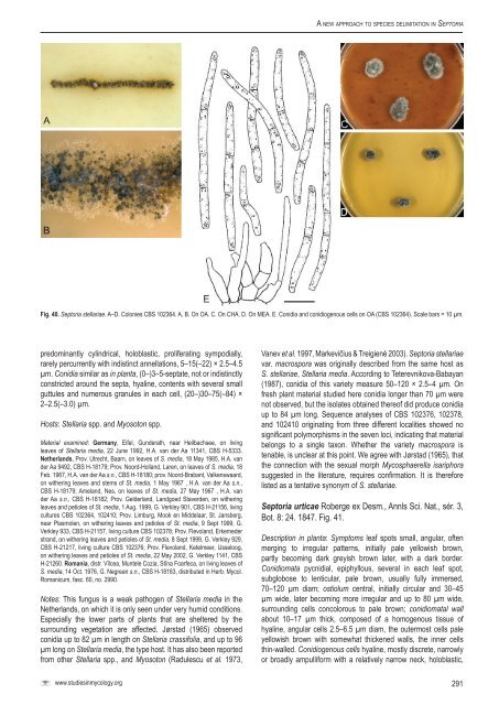
A <strong>new</strong> <strong>approach</strong> <strong>to</strong> <strong>species</strong> <strong>delimitation</strong> <strong>in</strong> Sep<strong>to</strong>riaFig. 40. Sep<strong>to</strong>ria stellariae. A–D. Colonies <strong>CBS</strong> 102364. A, B. On OA. C. On CHA. D. On MEA. E. Conidia and conidiogenous cells on OA (<strong>CBS</strong> 102364). Scale bars = 10 µm.predom<strong>in</strong>antly cyl<strong>in</strong>drical, holoblastic, proliferat<strong>in</strong>g sympodially,rarely percurrently with <strong>in</strong>dist<strong>in</strong>ct annellations, 5–15(–22) × 2.5–4.5µm. Conidia similar as <strong>in</strong> planta, (0–)3–5-septate, not or <strong>in</strong>dist<strong>in</strong>ctlyconstricted around the septa, hyal<strong>in</strong>e, contents with several smallguttules and numerous granules <strong>in</strong> each cell, (20–)30–75(–84) ×2–2.5(–3.0) µm.Hosts: Stellaria spp. and Myoso<strong>to</strong>n spp.Material exam<strong>in</strong>ed: Germany, Eifel, Gunderath, near Heilbachsee, on liv<strong>in</strong>gleaves of Stellaria media, 22 June 1992, H.A. van der Aa 11341, <strong>CBS</strong> H-5333.Netherlands, Prov. Utrecht, Baarn, on leaves of S. media, 18 May 1985, H.A. vander Aa 9492, <strong>CBS</strong> H-18179; Prov. Noord-Holland, Laren, on leaves of S. media, 18Feb. 1967, H.A. van der Aa s.n., <strong>CBS</strong> H-18180; prov. Noord-Brabant, Valkenswaard,on wither<strong>in</strong>g leaves and stems of St. media, 1 May 1967 , H.A. van der Aa s.n.,<strong>CBS</strong> H-18179; Ameland, Nes, on leaves of St. media, 27 May 1967 , H.A. vander Aa s.n., <strong>CBS</strong> H-18182; Prov. Gelderland, Landgoed Staverden, on wither<strong>in</strong>gleaves and petioles of St. media, 1 Aug. 1999, G. Verkley 901, <strong>CBS</strong> H-21156, liv<strong>in</strong>gcultures <strong>CBS</strong> 102364, 102410; Prov. Limburg, Mook en Middelaar, St. Jansberg,near Plasmolen, on wither<strong>in</strong>g leaves and petioles of St. media, 9 Sept 1999, G.Verkley 933, <strong>CBS</strong> H-21157, liv<strong>in</strong>g culture <strong>CBS</strong> 102378; Prov. Flevoland, Erkemederstrand, on wither<strong>in</strong>g leaves and petioles of St. media, 8 Sept 1999, G. Verkley 929,<strong>CBS</strong> H-21217, liv<strong>in</strong>g culture <strong>CBS</strong> 102376; Prov. Flevoland, Ketelmeer, IJsseloog,on wither<strong>in</strong>g leaves and petioles of St. media, 22 May 2002, G. Verkley 1141, <strong>CBS</strong>H-21260. Romania, distr. Vîlcea, Muntele Cozia, Stîna Foarfeca, on liv<strong>in</strong>g leaves ofS. media, 14 Oct. 1976, G. Negrean s.n., <strong>CBS</strong> H-18183, distributed <strong>in</strong> Herb. Mycol.Romanicum, fasc. 60, no. 2990.Notes: This fungus is a weak pathogen of Stellaria media <strong>in</strong> theNetherlands, on which it is only seen under very humid conditions.Especially the lower parts of plants that are sheltered by thesurround<strong>in</strong>g vegetation are affected. Jørstad (1965) observedconidia up <strong>to</strong> 82 µm <strong>in</strong> length on Stellaria crassifolia, and up <strong>to</strong> 96µm long on Stellaria media, the type host. It has also been reportedfrom other Stellaria spp., and Myoso<strong>to</strong>n (Radulescu et al. 1973,Vanev et al. 1997, Markevičius & Treigienė 2003). Sep<strong>to</strong>ria stellariaevar. macrospora was orig<strong>in</strong>ally described from the same host asS. stellariae, Stellaria media. Accord<strong>in</strong>g <strong>to</strong> Teterevnikova-Babayan(1987), conidia of this variety measure 50–120 × 2.5–4 µm. Onfresh plant material studied here conidia longer than 70 µm werenot observed, but the isolates obta<strong>in</strong>ed thereof did produce conidiaup <strong>to</strong> 84 µm long. Sequence analyses of <strong>CBS</strong> 102376, 102378,and 102410 orig<strong>in</strong>at<strong>in</strong>g from three different localities showed nosignificant polymorphisms <strong>in</strong> the seven loci, <strong>in</strong>dicat<strong>in</strong>g that materialbelongs <strong>to</strong> a s<strong>in</strong>gle taxon. Whether the variety macrospora istenable, is unclear at this po<strong>in</strong>t. We agree with Jørstad (1965), thatthe connection with the sexual morph Mycosphaerella isariphorasuggested <strong>in</strong> the literature, requires confirmation. It is thereforelisted as a tentative synonym of S. stellariae.Sep<strong>to</strong>ria urticae Roberge ex Desm., Annls Sci. Nat., sér. 3,Bot. 8: 24. 1847. Fig. 41.Description <strong>in</strong> planta: Symp<strong>to</strong>ms leaf spots small, angular, oftenmerg<strong>in</strong>g <strong>to</strong> irregular patterns, <strong>in</strong>itially pale yellowish brown,partly becom<strong>in</strong>g dark greyish brown later, with a dark border.Conidiomata pycnidial, epiphyllous, several <strong>in</strong> each leaf spot,subglobose <strong>to</strong> lenticular, pale brown, usually fully immersed,70–120 µm diam; ostiolum central, <strong>in</strong>itially circular and 30–45µm wide, later becom<strong>in</strong>g more irregular and up <strong>to</strong> 80 µm wide,surround<strong>in</strong>g cells concolorous <strong>to</strong> pale brown; conidiomatal wallabout 10–17 µm thick, composed of a homogenous tissue ofhyal<strong>in</strong>e, angular cells 2.5–6.5 µm diam, the outermost cells paleyellowish brown with somewhat thickened walls, the <strong>in</strong>ner cellsth<strong>in</strong>-walled. Conidiogenous cells hyal<strong>in</strong>e, mostly discrete, narrowlyor broadly ampulliform with a relatively narrow neck, holoblastic,www.studies<strong>in</strong>mycology.org291