

A new approach to species delimitation in Septoria - CBS - KNAW
A new approach to species delimitation in Septoria - CBS - KNAW
A new approach to species delimitation in Septoria - CBS - KNAW

You also want an ePaper? Increase the reach of your titles
YUMPU automatically turns print PDFs into web optimized ePapers that Google loves.
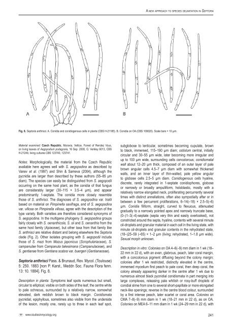
A <strong>new</strong> <strong>approach</strong> <strong>to</strong> <strong>species</strong> <strong>delimitation</strong> <strong>in</strong> Sep<strong>to</strong>riaFig. 8. Sep<strong>to</strong>ria anthrisci. A. Conidia and conidiogenous cells <strong>in</strong> planta (<strong>CBS</strong> H-21185). B. Conidia on OA (<strong>CBS</strong> 109020). Scale bars = 10 µm.Material exam<strong>in</strong>ed: Czech Republic, Moravia, Veltice, Forest of Rendez Vous,on liv<strong>in</strong>g leaves of Aegopodium podagraria, 16 Sep. 2008, G. Verkley 6013, <strong>CBS</strong>H-21249, liv<strong>in</strong>g cultures <strong>CBS</strong> 123740, 123741.Notes: Morphologically, the material from the Czech Republicavailable here agrees well with S. aegopod<strong>in</strong>a as described byVanev et al. (1997) and Sh<strong>in</strong> & Sameva (2004), although thepycnidia are larger than described by these authors (55–85 μmdiam). The <strong>species</strong> can easily be dist<strong>in</strong>guished from S. aegopodiioccurr<strong>in</strong>g on the same host plant, as the conidia of that fungusare considerably larger (30–115 × 3.5–4 µm), and appearpredom<strong>in</strong>antly 1-septate. The conidia more closely resemblethose of S. anthrisci. The diagnoses of S. aegopod<strong>in</strong>a var. trailiibased on material on Pimp<strong>in</strong>ella saxifraga, and of S. aegopod<strong>in</strong>avar. villosa on Pimp<strong>in</strong>ella villosa, agree with the description of thetype variety. Both varieties are therefore considered synonyms ofS. aegopod<strong>in</strong>a. In the multigene phylogeny S. aegopod<strong>in</strong>a groupsfairly closely with S. oenanthicola, S. sii and S. oenanthis from thesame host family (Apiaceae), but other taxa from that family likeS. anthrisci are relative distant and belong elsewhere the Sep<strong>to</strong>riaclade (Fig. 2). Other isolates group<strong>in</strong>g with S. aegopodii <strong>in</strong>cludethose of S. mazi from Mazus japonicus (Scrophulariaceae), S.campanulae from Campanula takesimana (Campanulaceae), andS. gentianae from Gentiana scabra var. buergeri (Gentianaceae).Sep<strong>to</strong>ria anthrisci Pass. & Brunaud, Rev. Mycol. (Toulouse)5: 250. 1883 [non P. Karst., Meddn Soc. Fauna Flora fenn.13: 10. 1884]. Fig. 8.Description <strong>in</strong> planta: Symp<strong>to</strong>ms leaf spots numerous but small,circular <strong>to</strong> elliptical, visible on both sides of the leaf, the centre white<strong>to</strong> pale ochreous, surrounded by a relatively narrow, somewhatelevated, dark reddish brown <strong>to</strong> black marg<strong>in</strong>. Conidiomatapycnidial, epiphyllous, sometimes also visible from the undersideof the lesion, mostly one, rarely up <strong>to</strong> three <strong>in</strong> each leaf spot,subglobose <strong>to</strong> lenticular, sometimes becom<strong>in</strong>g cupulate, brown<strong>to</strong> black, immersed, 115–190 µm diam; ostiolum central, <strong>in</strong>itiallycircular and 30–55 µm wide, later becom<strong>in</strong>g more irregular andup <strong>to</strong> 100 µm wide, surround<strong>in</strong>g cells concolorous; conidiomatalwall about 12–20 µm thick, composed of an outer layer of palebrown angular cells 4.5–7 µm diam with somewhat thickenedwalls, and an <strong>in</strong>ner layer of th<strong>in</strong>-walled, pale yellow angular<strong>to</strong> globose cells 2.5–5 µm diam. Conidiogenous cells hyal<strong>in</strong>e,discrete, rarely <strong>in</strong>tegrated <strong>in</strong> 1-septate conidiophores, globoseor narrowly or broadly ampulliform, holoblastic, mostly with arelatively narrow elongated neck, proliferat<strong>in</strong>g percurrently severaltimes with dist<strong>in</strong>ct annellations, often also sympodially after or <strong>in</strong>between a few percurrent proliferations, 6–14(–18) × 2.5–5(–6)µm. Conidia filiform, straight, curved <strong>to</strong> flexuous, attenuatedgradually <strong>to</strong> a narrowly po<strong>in</strong>ted apex and narrowly truncate base,(0–)1–3(–4)-septate (septa very th<strong>in</strong> and easily overlooked), notconstricted around the septa, hyal<strong>in</strong>e, contents with several m<strong>in</strong>uteoil-droplets and granular material <strong>in</strong> each cell <strong>in</strong> the liv<strong>in</strong>g state, withm<strong>in</strong>ute oil-droplets and granular contents <strong>in</strong> the rehydrated state,(18–)25–59 (–65) × 1–2 µm (liv<strong>in</strong>g; rehydrated, 1–1.8 µm wide).Sexual morph unknown.Description <strong>in</strong> vitro: Colonies on OA 4–6(–9) mm diam <strong>in</strong> 1 wk (18–22 mm <strong>in</strong> 22 d), with an even, glabrous, peach, later coral marg<strong>in</strong>,with a concolorous pigment diffus<strong>in</strong>g beyond the colony marg<strong>in</strong>;colonies after 1 wk restricted, dist<strong>in</strong>ctly elevated <strong>in</strong> the centre,immersed mycelium first peach <strong>to</strong> pale coral, then deep coral, thecolony already appear<strong>in</strong>g darker <strong>in</strong> the centre after 1 wk due <strong>to</strong>numerous almost black pycnidial conidiomata <strong>in</strong> part merg<strong>in</strong>g <strong>in</strong><strong>to</strong>large complexes, releas<strong>in</strong>g pale whitish or rosy-buff droplets ofconidial slime from one <strong>to</strong> several short-papillate or more elongatedneck-like open<strong>in</strong>gs; reverse <strong>in</strong> the centre blood colour, surroundedby a first <strong>in</strong>tense peach, later scarlet or coral area. Colonies onCMA 7–8(–9) mm diam <strong>in</strong> 1 wk (18–21 mm <strong>in</strong> 22 d), as on OA.Colonies on MEA 6–11 mm diam <strong>in</strong> 1 wk (24–29 mm <strong>in</strong> 22 d), withwww.studies<strong>in</strong>mycology.org241


