Verkley et al.<strong>to</strong> very dark dull green, with numerous dark, radiat<strong>in</strong>g hyphae,almost entirely glabrous, few tufts of greyish aerial mycelium<strong>in</strong> the centre; numerous scattered s<strong>in</strong>gle or complex pycnidialconidiomata developed already after 1 wk, with a s<strong>in</strong>gle ostiole orseveral papillate or rostrate open<strong>in</strong>gs, from which pale rosy-buffdroplets of conidial slime are released; reverse concolourous.Colonies on CMA 16–18(–20) mm diam <strong>in</strong> 3 wk (38–50 mm <strong>in</strong> 6wk), as on OA. Colonies on MEA 9–12(–14) mm diam <strong>in</strong> 3 wk (27–39 mm <strong>in</strong> 6 wk), with an even <strong>to</strong> slightly ruffled buff marg<strong>in</strong>; coloniesrestricted, conical and up <strong>to</strong> 3 mm high after 3 wk, immersedmycelium near the marg<strong>in</strong> grey-olivaceous, but most of the colonysurface iron grey <strong>to</strong> greenish black, the outer areas mostly coveredby a low but dense, f<strong>in</strong>ely felted, grey aerial mycelium, the centrealmost glabrous; superficial semi-immersed conidiomata releas<strong>in</strong>gpale whitish droplets of conidial slime after 2–3 wk; reverse mostlydark slate blue with olivaceous areas. Colonies on CHA 16–22 mmdiam <strong>in</strong> 3 wk (39–46 mm <strong>in</strong> 6 wk), as on MEA, but conidiomatamore numerous, releas<strong>in</strong>g pale whitish <strong>to</strong> pale rosy-buff droplets orcirrhi of conidial slime, and reverse with a brown-v<strong>in</strong>aceous t<strong>in</strong>ge.Conidiomata as <strong>in</strong> planta, pycnidial with a s<strong>in</strong>gle ostiolum,dark brown <strong>to</strong> black, rarely merged <strong>in</strong><strong>to</strong> complex fruitbodies;conidiogenous cells as <strong>in</strong> planta, but larger and more often<strong>in</strong>tegrated <strong>in</strong> 1–3-septate conidiophores, 10–15(–23) × 3–6(–7)µm; conidia as <strong>in</strong> planta, but longer, 36–78(–90) × (1.6–)1.7–2.2µm, contents several oil-droplets <strong>in</strong> each cell.Hosts: Matricaria spp.Material exam<strong>in</strong>ed: Netherlands, prov. Limburg, Zuid-Limburg, along roadside nearSavelsbos, on liv<strong>in</strong>g leaves of Matricaria discoidea (= M. matricarioides), 28 June2000, G. Verkley 960, <strong>CBS</strong> H-21228, liv<strong>in</strong>g cultures <strong>CBS</strong> 109000, 109001. Romania,Suceava, Siret, on leaves of M. discoidea, 7 July 1969, distributed <strong>in</strong> Contant<strong>in</strong>escu& Negrean, Herb. mycol. Romanicum Fasc. 40, no. 199, <strong>CBS</strong> H-18115.Notes: The phylogenetic analyses <strong>in</strong>dicate that S. matricariae isclosest <strong>to</strong> S. lamiicola, yet rather distant from other Sep<strong>to</strong>ria occurr<strong>in</strong>gon Asteraceae. The <strong>in</strong>def<strong>in</strong>ite lesions caused by this <strong>species</strong>are rem<strong>in</strong>iscent of those developed by S. stellariae on Stellariamedia. The leaves seem <strong>to</strong> whither more rapidly and pycnidiadevelop soon after discolouration of the leaf tissues starts. Stemsare not affected. In the orig<strong>in</strong>al diagnosis of S. matricariae, basedon material from Matricaria discoidea <strong>in</strong> Hungary, the conidia aredescribed as cont<strong>in</strong>uous and 40–60 × 2–2.5 µm. The Dutch and alsothe Romanian material studied here conta<strong>in</strong> conidia with mostly 1–3septa, but otherwise agree well with Hollós’ description of the type.Accord<strong>in</strong>g <strong>to</strong> Radulescu et al. (1973) the conidia <strong>in</strong> material from thesame host plant are also cont<strong>in</strong>uous, measur<strong>in</strong>g 25–50 × 1.5–2 µm.As <strong>in</strong> several other Sep<strong>to</strong>ria spp., the septa <strong>in</strong> S. matricariae are noteasy <strong>to</strong> observe, and Hollós and others may have overlooked them.Sydow described a Sep<strong>to</strong>ria under the same name fromMatricaria chamomilla <strong>in</strong> Germany with multiseptate conidia 30–60× 1–1.5 µm. The name he proposed was illegitimate because it isa later homonym of S. matricariae Hollós, as is also S. matricariaeCejp et al.. Sep<strong>to</strong>ria chamomillae was also described from M.chamomillae <strong>in</strong> Belgium and has 3–5-septate conidia 35–52 × 1–2µm. Although we have not seen the types of either of these names,we consider them tentatively as synonyms of S. matricariae.Sep<strong>to</strong>ria melissae Desm., Annls Sci. Nat., sér. 3, Bot., 20:87. 1853. Fig. 29.≡ Phloeospora melissae (Desm.) Parisi, Bull. Bot. R. Univ. Napoli 6: 292.1921.Description <strong>in</strong> vitro: Colonies on OA 12–13 mm diam <strong>in</strong> 2 wk, withan even <strong>to</strong> slightly ruffled, mostly colourless marg<strong>in</strong>; coloniesrestricted <strong>to</strong> spread<strong>in</strong>g, somewhat elevated <strong>in</strong> the centre, immersedmycelium greenish black, with greenish hyphal strands radiat<strong>in</strong>g <strong>in</strong><strong>to</strong>or even beyond the colourless marg<strong>in</strong>, the surface mostly glabrousor provided with very diffuse, f<strong>in</strong>ely felted, grey aerial hyphae,the elevations <strong>in</strong> the centre bear<strong>in</strong>g tufts of more well-developed,grey aerial mycelium; conidiomata develop<strong>in</strong>g mostly <strong>in</strong> the centreimmersed or on the agar surface, releas<strong>in</strong>g pale rosy <strong>to</strong> rosy-buffconidial slime. No diffus<strong>in</strong>g pigment observed. Colonies on MEA5–7(–9) mm diam <strong>in</strong> 2 wk, with a slightly ruffled marg<strong>in</strong>; coloniesrestricted, pustulate with cerebriform elevations <strong>in</strong> the centre, thesurface black, covered by a diffuse <strong>to</strong> dense mat of f<strong>in</strong>ely felted,mostly grey aerial mycelium; reverse very dark brown-v<strong>in</strong>aceous.Conidiomata sparsely develop<strong>in</strong>g on the colony surface, releas<strong>in</strong>gdirty reddish brown conidial slime. A very fa<strong>in</strong>t pigment is visiblearound the colony.Conidiogenous cells (OA) globose <strong>to</strong> ampulliform, holoblastic,hyal<strong>in</strong>e, discrete or <strong>in</strong>tegrated <strong>in</strong> 1(–2)-septate conidiophores,proliferat<strong>in</strong>g sympodially, percurrent proliferation not observed,4–10 × 3–5 µm. Conidia filiform, straight <strong>to</strong> flexuous, weakly <strong>to</strong>more strongly curved, attenuated gradually <strong>to</strong> a narrowly rounded,typically po<strong>in</strong>ted apex, attenuated gradually <strong>to</strong> a narrowly truncate<strong>to</strong> somewhat rounded base, hyal<strong>in</strong>e, with f<strong>in</strong>e granular material andm<strong>in</strong>ute oil-droplets, (0–)3(–5)-septate, (22–)30–50(–61) × 1.5–2µm. Sexual morph unknown.Host: Melissa offic<strong>in</strong>alis.Material exam<strong>in</strong>ed: Netherlands, Baarn, garden Eemnesserweg, on liv<strong>in</strong>g leavesof Melissa offic<strong>in</strong>alis, 11 Sep. 2000, H.A. van der Aa s.n. (G. Verkley 1073), <strong>CBS</strong>H-21169, liv<strong>in</strong>g cultures <strong>CBS</strong> 109096, 109097.Notes: This <strong>species</strong> is the only Sep<strong>to</strong>ria described from the genusMelissa. The type material orig<strong>in</strong>ates from Melissa offic<strong>in</strong>alis <strong>in</strong>France (not seen). Accord<strong>in</strong>g <strong>to</strong> the short orig<strong>in</strong>al diagnosis, S.melissae produces conidia 30 × 1.6 µm, and no septa were reported.Radulescu et al. (1973) described the conidia as cont<strong>in</strong>uous or with1–3 septa, 25–38 × 1.6 µm. These measurements agree quitewell with those given by Teterevnikova-Babayan (1987; 28–38× 1.5 µm), but Vanev et al. (1997) gave a much wider range ofmeasurements, 20.5–58 × 1.5–2.2 µm (septa 2–5). Genetically<strong>CBS</strong> 109097 is very closely related <strong>to</strong> S. galeopsidis, but a 5 bp<strong>in</strong>sertion found <strong>in</strong> the Btub gene is absent <strong>in</strong> all sequenced stra<strong>in</strong>s ofS. galeopsidis. Sep<strong>to</strong>ria melissae can furthermore be dist<strong>in</strong>guished<strong>in</strong> culture from S. galeopsidis by the narrower conidia on OA (1.5–2µm, <strong>in</strong> S. galeopsidis 2–2.5 µm), and the conidiogenous cells,which only proliferate sympodially and not percurrently.Sep<strong>to</strong>ria napelli Speg, Decades mycologicae italicae I-XII:no. 117. 1879; Atti Soc. crit<strong>to</strong>g. ital., Ser. 2, 3: 69. 1880. Fig.30.≡ Rhabdospora napelli (Speg.) Petr., Sydowia 11: 376. 1957[misapplication].Description <strong>in</strong> planta: Symp<strong>to</strong>ms leaf spots hologenous, circular <strong>to</strong>irregular, s<strong>in</strong>gle, white <strong>to</strong> pale greyish, surrounded by a first red,then black, relatively wide border, often completely blacken<strong>in</strong>g thenarrow leaflets. Conidiomata pycnidial, epiphyllous, rarely alsohypohyllous, conspicuous, one <strong>to</strong> many <strong>in</strong> each leaf spot, globose<strong>to</strong> subglobose, black, semi-immersed, 100–150(–200) µm diam;ostiolum central, circular, <strong>in</strong>itially 15–25 µm wide, later open<strong>in</strong>g272
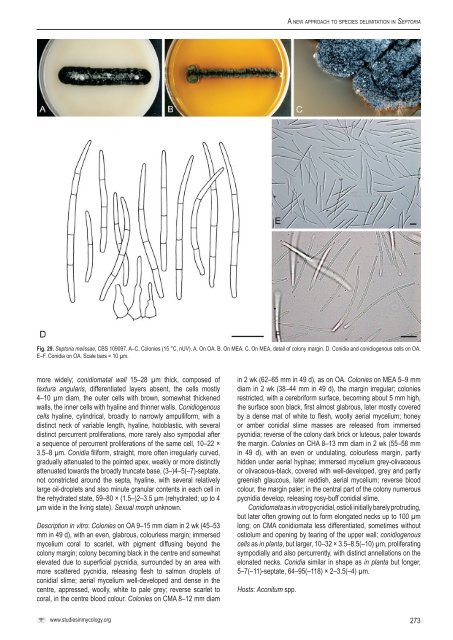
A <strong>new</strong> <strong>approach</strong> <strong>to</strong> <strong>species</strong> <strong>delimitation</strong> <strong>in</strong> Sep<strong>to</strong>riaFig. 29. Sep<strong>to</strong>ria melissae, <strong>CBS</strong> 109097. A–C. Colonies (15 °C, nUV). A. On OA. B. On MEA. C. On MEA, detail of colony marg<strong>in</strong>. D. Conidia and conidiogenous cells on OA.E–F. Conidia on OA. Scale bars = 10 µm.more widely; conidiomatal wall 15–28 µm thick, composed oftextura angularis, differentiated layers absent, the cells mostly4–10 µm diam, the outer cells with brown, somewhat thickenedwalls, the <strong>in</strong>ner cells with hyal<strong>in</strong>e and th<strong>in</strong>ner walls. Conidiogenouscells hyal<strong>in</strong>e, cyl<strong>in</strong>drical, broadly <strong>to</strong> narrowly ampulliform, with adist<strong>in</strong>ct neck of variable length, hyal<strong>in</strong>e, holoblastic, with severaldist<strong>in</strong>ct percurrent proliferations, more rarely also sympodial aftera sequence of percurrent proliferations of the same cell, 10–22 ×3.5–8 µm. Conidia filiform, straight, more often irregularly curved,gradually attenuated <strong>to</strong> the po<strong>in</strong>ted apex, weakly or more dist<strong>in</strong>ctlyattenuated <strong>to</strong>wards the broadly truncate base, (3–)4–5(–7)-septate,not constricted around the septa, hyal<strong>in</strong>e, with several relativelylarge oil-droplets and also m<strong>in</strong>ute granular contents <strong>in</strong> each cell <strong>in</strong>the rehydrated state, 59–80 × (1.5–)2–3.5 µm (rehydrated; up <strong>to</strong> 4µm wide <strong>in</strong> the liv<strong>in</strong>g state). Sexual morph unknown.Description <strong>in</strong> vitro: Colonies on OA 9–15 mm diam <strong>in</strong> 2 wk (45–53mm <strong>in</strong> 49 d), with an even, glabrous, colourless marg<strong>in</strong>; immersedmycelium coral <strong>to</strong> scarlet, with pigment diffus<strong>in</strong>g beyond thecolony marg<strong>in</strong>; colony becom<strong>in</strong>g black <strong>in</strong> the centre and somewhatelevated due <strong>to</strong> superficial pycnidia, surrounded by an area withmore scattered pycnidia, releas<strong>in</strong>g flesh <strong>to</strong> salmon droplets ofconidial slime; aerial mycelium well-developed and dense <strong>in</strong> thecentre, appressed, woolly, white <strong>to</strong> pale grey; reverse scarlet <strong>to</strong>coral, <strong>in</strong> the centre blood colour. Colonies on CMA 8–12 mm diam<strong>in</strong> 2 wk (62–65 mm <strong>in</strong> 49 d), as on OA. Colonies on MEA 5–9 mmdiam <strong>in</strong> 2 wk (38–44 mm <strong>in</strong> 49 d), the marg<strong>in</strong> irregular; coloniesrestricted, with a cerebriform surface, becom<strong>in</strong>g about 5 mm high,the surface soon black, first almost glabrous, later mostly coveredby a dense mat of white <strong>to</strong> flesh, woolly aerial mycelium; honeyor amber conidial slime masses are released from immersedpycnidia; reverse of the colony dark brick or luteous, paler <strong>to</strong>wardsthe marg<strong>in</strong>. Colonies on CHA 8–13 mm diam <strong>in</strong> 2 wk (55–58 mm<strong>in</strong> 49 d), with an even or undulat<strong>in</strong>g, colourless marg<strong>in</strong>, partlyhidden under aerial hyphae; immersed mycelium grey-olivaceousor olivaceous-black, covered with well-developed, grey and partlygreenish glaucous, later reddish, aerial mycelium; reverse bloodcolour, the marg<strong>in</strong> paler; <strong>in</strong> the central part of the colony numerouspycnidia develop, releas<strong>in</strong>g rosy-buff conidial slime.Conidiomata as <strong>in</strong> vitro pycnidial, ostioli <strong>in</strong>itially barely protrud<strong>in</strong>g,but later often grow<strong>in</strong>g out <strong>to</strong> form elongated necks up <strong>to</strong> 100 µmlong; on CMA conidiomata less differentiated, sometimes withou<strong>to</strong>stiolum and open<strong>in</strong>g by tear<strong>in</strong>g of the upper wall; conidiogenouscells as <strong>in</strong> planta, but larger, 10–32 × 3.5–8.5(–10) µm, proliferat<strong>in</strong>gsympodially and also percurrently, with dist<strong>in</strong>ct annellations on theelonated necks. Conidia similar <strong>in</strong> shape as <strong>in</strong> planta but longer,5–7(–11)-septate, 64–95(–118) × 2–3.5(–4) µm.Hosts: Aconitum spp.www.studies<strong>in</strong>mycology.org273