

A new approach to species delimitation in Septoria - CBS - KNAW
A new approach to species delimitation in Septoria - CBS - KNAW
A new approach to species delimitation in Septoria - CBS - KNAW

Create successful ePaper yourself
Turn your PDF publications into a flip-book with our unique Google optimized e-Paper software.
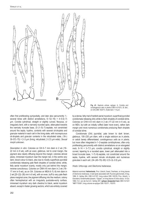
Verkley et al.Fig. 41. Sep<strong>to</strong>ria urticae, epitype. A. Conidia andconidiogenous cells <strong>in</strong> planta (<strong>CBS</strong> H-21221). B. Ibid.,on OA (<strong>CBS</strong> 102371). Scale bars = 10 µm.often first proliferat<strong>in</strong>g sympodially, and later also percurrently 1–several times with dist<strong>in</strong>ct annellations, 6–12(–16) × 4–5.5(–7)µm. Conidia cyl<strong>in</strong>drical, straight or slightly curved, flexuous, orirregularly bent, with a narrowly rounded apex, attenuated <strong>to</strong>wardsthe narrowly truncate base, (0–)1–5(–7)-septate, not constrictedaround the septa, hyal<strong>in</strong>e, contents with several oil-droplets andgranular material <strong>in</strong> each cell <strong>in</strong> the liv<strong>in</strong>g state, with <strong>in</strong>conspicuousoil-droplets and granular contents <strong>in</strong> the rehydrated state, (18–)30–57(–75) × 2– 3 µm (liv<strong>in</strong>g; rehydrated, 2–2.5 µm wide). Sexualmorph unknown.Description <strong>in</strong> vitro: Colonies on OA 6–7 mm diam <strong>in</strong> 2 wk (19–22 mm <strong>in</strong> 6 wk), with an even, glabrous, red <strong>to</strong> coral marg<strong>in</strong>, thepigment also clearly diffus<strong>in</strong>g beyond the marg<strong>in</strong>; colonies almostplane, immersed mycelium near the marg<strong>in</strong> red, <strong>in</strong> the centre verydark, blood colour <strong>to</strong> black, also due <strong>to</strong> mostly superficial pycnidialconidiomata releas<strong>in</strong>g pale flesh droplets of conidial slime; white,felty aerial mycelium scanty, mostly only just beh<strong>in</strong>d the marg<strong>in</strong>;reverse concolorous. Colonies on CMA 4-6 mm diam <strong>in</strong> 2 wk (16–17 mm <strong>in</strong> 6 wk), as on OA. Colonies on MEA 6–7(–9) mm diam <strong>in</strong>2 wk [20–22(–28) mm <strong>in</strong> 6 wk], with an even, buff <strong>to</strong> very pale fleshplane marg<strong>in</strong>al zone; the pigment diffus<strong>in</strong>g <strong>in</strong><strong>to</strong> the medium; colonyoften hemispherical with an irregularly pustulate-worty surface,immersed mycelium very dark chestnut <strong>to</strong> black, aerial myceliumabsent, except <strong>in</strong> faster grow<strong>in</strong>g sec<strong>to</strong>rs, which are entirely coveredby a dense, felty mat of reddish aerial mycelium; superficial pycnidialconidiomata releas<strong>in</strong>g dirty white <strong>to</strong> flesh droplets of conidial slime.Colonies on CHA 4–6 mm diam <strong>in</strong> 2 wk (17–22 mm <strong>in</strong> 6 wk), ason MEA, but with an <strong>in</strong>itially ruffled (later more even), rather darkmarg<strong>in</strong> and more numerous conidiomata produc<strong>in</strong>g flesh dropletsof conidial slime.Conidiomata (OA) pycnidial, pale brown <strong>to</strong> dark brown,glabrous, 100–230 µm diam, with a s<strong>in</strong>gle ostiolum as <strong>in</strong> planta,or ostioli barely differentiated; conidiogenous cells as <strong>in</strong> planta,but more often <strong>in</strong>tegrated <strong>in</strong> 1–2-septate conidiophores, often onlyproliferat<strong>in</strong>g percurrently with dist<strong>in</strong>ct annellations on an elongatedneck, 6–14 × 3–7.5 µm; conidia cyl<strong>in</strong>drical, straight or slightlycurved, taper<strong>in</strong>g <strong>to</strong> a rounded apex, lower part attenuated <strong>in</strong><strong>to</strong> abroad truncate base, 1–7(–9)-septate, not constricted around thesepta, hyal<strong>in</strong>e, with several m<strong>in</strong>ute oil-droplets and numerousgranulae <strong>in</strong> each cell, (34–)40–70(–90) ×2.5–3(–3.5) µm.Hosts: Urtica spp. and Glechoma hederacea.Material exam<strong>in</strong>ed: Netherlands, Prov. Utrecht, Soest, Overhees, on liv<strong>in</strong>g leavesof Glechoma hederacea, <strong>in</strong> leaf spots associated with Pucc<strong>in</strong>ia glechomatis, 8 Aug.1999, G. Verkley 904, <strong>CBS</strong> H-21197, liv<strong>in</strong>g culture <strong>CBS</strong> 102316; Prov. Utrecht, ‘sGraveland, Kortenhoefse plassen, “Oppad”, on liv<strong>in</strong>g leaves of Urtica dioica, 14 Oct.1999, H.A. van der Aa & G. Verkley 947, epitype designated here <strong>CBS</strong> H-21221“MBT175359”, liv<strong>in</strong>g cultures ex-epitype <strong>CBS</strong> 102371, 102375.292


