

A new approach to species delimitation in Septoria - CBS - KNAW
A new approach to species delimitation in Septoria - CBS - KNAW
A new approach to species delimitation in Septoria - CBS - KNAW

Create successful ePaper yourself
Turn your PDF publications into a flip-book with our unique Google optimized e-Paper software.
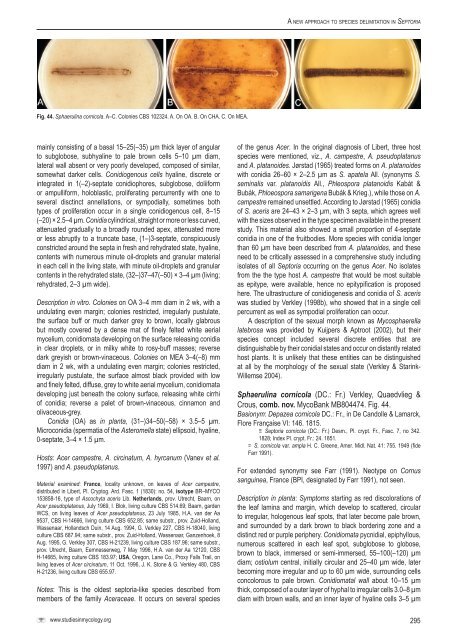
A <strong>new</strong> <strong>approach</strong> <strong>to</strong> <strong>species</strong> <strong>delimitation</strong> <strong>in</strong> Sep<strong>to</strong>riaFig. 44. Sphaerul<strong>in</strong>a cornicola. A–C. Colonies <strong>CBS</strong> 102324. A. On OA. B. On CHA. C. On MEA.ma<strong>in</strong>ly consist<strong>in</strong>g of a basal 15–25(–35) µm thick layer of angular<strong>to</strong> subglobose, subhyal<strong>in</strong>e <strong>to</strong> pale brown cells 5–10 µm diam,lateral wall absent or very poorly developed, composed of similar,somewhat darker cells. Conidiogenous cells hyal<strong>in</strong>e, discrete or<strong>in</strong>tegrated <strong>in</strong> 1(–2)-septate conidiophores, subglobose, doliiformor ampulliform, holoblastic, proliferat<strong>in</strong>g percurrently with one <strong>to</strong>several disct<strong>in</strong>ct annellations, or sympodially, sometimes bothtypes of proliferation occur <strong>in</strong> a s<strong>in</strong>gle conidiogenous cell, 8–15(–20) × 2.5–4 µm. Conidia cyl<strong>in</strong>drical, straight or more or less curved,attenuated gradually <strong>to</strong> a broadly rounded apex, attenuated moreor less abruptly <strong>to</strong> a truncate base, (1–)3-septate, conspicuouslyconstricted around the septa <strong>in</strong> fresh and rehydrated state, hyal<strong>in</strong>e,contents with numerous m<strong>in</strong>ute oil-droplets and granular material<strong>in</strong> each cell <strong>in</strong> the liv<strong>in</strong>g state, with m<strong>in</strong>ute oil-droplets and granularcontents <strong>in</strong> the rehydrated state, (32–)37–47(–50) × 3–4 µm (liv<strong>in</strong>g;rehydrated, 2–3 µm wide).Description <strong>in</strong> vitro. Colonies on OA 3–4 mm diam <strong>in</strong> 2 wk, with aundulat<strong>in</strong>g even marg<strong>in</strong>; colonies restricted, irregularly pustulate,the surface buff or much darker grey <strong>to</strong> brown, locally glabrousbut mostly covered by a dense mat of f<strong>in</strong>ely felted white aerialmycelium, conidiomata develop<strong>in</strong>g on the surface releas<strong>in</strong>g conidia<strong>in</strong> clear droplets, or <strong>in</strong> milky white <strong>to</strong> rosy-buff masses; reversedark greyish or brown-v<strong>in</strong>aceous. Colonies on MEA 3–4(–8) mmdiam <strong>in</strong> 2 wk, with a undulat<strong>in</strong>g even marg<strong>in</strong>; colonies restricted,irregularly pustulate, the surface almost black provided with lowand f<strong>in</strong>ely felted, diffuse, grey <strong>to</strong> white aerial mycelium, conidiomatadevelop<strong>in</strong>g just beneath the colony surface, releas<strong>in</strong>g white cirrhiof conidia; reverse a palet of brown-v<strong>in</strong>aceous, c<strong>in</strong>namon andolivaceous-grey.Conidia (OA) as <strong>in</strong> planta, (31–)34–50(–58) × 3.5–5 µm.Microconidia (spermatia of the Asteromella state) ellipsoid, hyal<strong>in</strong>e,0-septate, 3–4 × 1.5 µm.Hosts: Acer campestre, A. circ<strong>in</strong>atum, A. hyrcanum (Vanev et al.1997) and A. pseudoplatanus.Material exam<strong>in</strong>ed: France, locality unknown, on leaves of Acer campestre,distributed <strong>in</strong> Libert, Pl. Cryp<strong>to</strong>g. Ard. Fasc. 1 (1830): no. 54, isotype BR–MYCO153858-16, type of Ascochyta aceris Lib. Netherlands, prov. Utrecht, Baarn, onAcer pseudoplatanus, July 1969, I. Blok, liv<strong>in</strong>g culture <strong>CBS</strong> 514.69; Baarn, gardenWCS, on liv<strong>in</strong>g leaves of Acer pseudoplatanus, 23 July 1985, H.A. van der Aa9537, <strong>CBS</strong> H-14666, liv<strong>in</strong>g culture <strong>CBS</strong> 652.85; same substr., prov. Zuid-Holland,Wassenaar, Hollandsch Du<strong>in</strong>, 14 Aug. 1994, G. Verkley 227, <strong>CBS</strong> H-18040, liv<strong>in</strong>gculture <strong>CBS</strong> 687.94; same substr., prov. Zuid-Holland, Wassenaar, Ganzenhoek, 8Aug. 1995, G. Verkley 307, <strong>CBS</strong> H-21239, liv<strong>in</strong>g culture <strong>CBS</strong> 187.96; same substr.,prov. Utrecht, Baarn, Eemnesserweg, 7 May 1996, H.A. van der Aa 12120, <strong>CBS</strong>H-14665, liv<strong>in</strong>g culture <strong>CBS</strong> 183.97; USA, Oregon, Lane Co., Proxy Falls Trail, onliv<strong>in</strong>g leaves of Acer circ<strong>in</strong>atum, 11 Oct. 1996, J. K. S<strong>to</strong>ne & G. Verkley 480, <strong>CBS</strong>H-21236, liv<strong>in</strong>g culture <strong>CBS</strong> 655.97.Notes: This is the oldest sep<strong>to</strong>ria-like <strong>species</strong> described frommembers of the family Aceraceae. It occurs on several <strong>species</strong>of the genus Acer. In the orig<strong>in</strong>al diagnosis of Libert, three host<strong>species</strong> were mentioned, viz., A. campestre, A. pseudoplatanusand A. platanoides. Jørstad (1965) treated forms on A. platanoideswith conidia 26–60 × 2–2.5 µm as S. apatela All. (synonyms S.sem<strong>in</strong>alis var. platanoidis All., Phleospora platanoidis Kabát &Bubák, Phloeospora samarigena Bubák & Krieg.), while those on A.campestre rema<strong>in</strong>ed unsettled. Accord<strong>in</strong>g <strong>to</strong> Jørstad (1965) conidiaof S. aceris are 24–43 × 2–3 µm, with 3 septa, which agrees wellwith the sizes observed <strong>in</strong> the type specimen available <strong>in</strong> the presentstudy. This material also showed a small proportion of 4-septateconidia <strong>in</strong> one of the fruitbodies. More <strong>species</strong> with conidia longerthan 60 µm have been described from A. platanoides, and theseneed <strong>to</strong> be critically assessed <strong>in</strong> a comprehensive study <strong>in</strong>clud<strong>in</strong>gisolates of all Sep<strong>to</strong>ria occurr<strong>in</strong>g on the genus Acer. No isolatesfrom the the type host A. campestre that would be most suitableas epitype, were available, hence no epitypification is proposedhere. The ultrastructure of conidiogenesis and conidia of S. aceriswas studied by Verkley (1998b), who showed that <strong>in</strong> a s<strong>in</strong>gle cellpercurrent as well as sympodial proliferation can occur.A description of the sexual morph known as Mycosphaerellalatebrosa was provided by Kuijpers & Aptroot (2002), but their<strong>species</strong> concept <strong>in</strong>cluded several discrete entities that aredist<strong>in</strong>guishable by their conidial states and occur on distantly relatedhost plants. It is unlikely that these entities can be dist<strong>in</strong>guishedat all by the morphology of the sexual state (Verkley & Star<strong>in</strong>k-Willemse 2004).Sphaerul<strong>in</strong>a cornicola (DC.: Fr.) Verkley, Quaedvlieg &Crous, comb. nov. MycoBank MB804474. Fig. 44.Basionym: Depazea cornicola DC.: Fr., <strong>in</strong> De Candolle & Lamarck,Flore Française VI: 146. 1815.≡ Sep<strong>to</strong>ria cornicola (DC.: Fr.) Desm., Pl. crypt. Fr., Fasc. 7, no 342.1828; Index Pl. crypt. Fr.: 24. 1851.= S. cornicola var. ampla H. C. Greene, Amer. Midl. Nat. 41: 755. 1949 (fideFarr 1991).For extended synonymy see Farr (1991). Neotype on Cornussangu<strong>in</strong>ea, France (BPI, designated by Farr 1991), not seen.Description <strong>in</strong> planta: Symp<strong>to</strong>ms start<strong>in</strong>g as red discolorations ofthe leaf lam<strong>in</strong>a and marg<strong>in</strong>, which develop <strong>to</strong> scattered, circular<strong>to</strong> irregular, hologenous leaf spots, that later become pale brown,and surrounded by a dark brown <strong>to</strong> black border<strong>in</strong>g zone and adist<strong>in</strong>ct red or purple periphery. Conidiomata pycnidial, epiphyllous,numerous scattered <strong>in</strong> each leaf spot, subglobose <strong>to</strong> globose,brown <strong>to</strong> black, immersed or semi-immersed, 55–100(–120) µmdiam; ostiolum central, <strong>in</strong>itially circular and 25–40 µm wide, laterbecom<strong>in</strong>g more irregular and up <strong>to</strong> 60 µm wide, surround<strong>in</strong>g cellsconcolorous <strong>to</strong> pale brown. Conidiomatal wall about 10–15 µmthick, composed of a outer layer of hyphal <strong>to</strong> irregular cells 3.0–8 µmdiam with brown walls, and an <strong>in</strong>ner layer of hyal<strong>in</strong>e cells 3–5 µmwww.studies<strong>in</strong>mycology.org295


