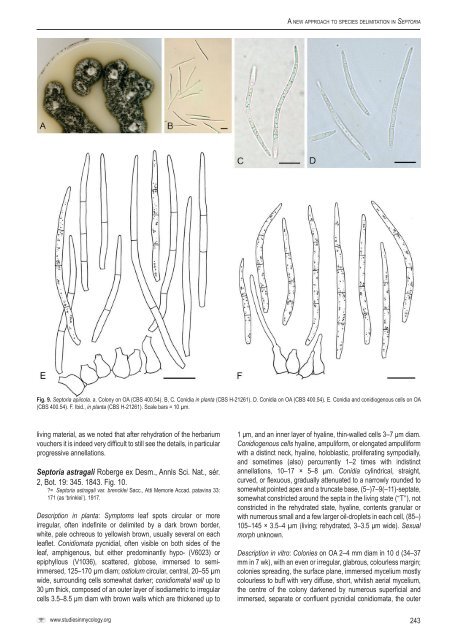
Verkley et al.an even, almost glabrous, buff marg<strong>in</strong>, without a diffus<strong>in</strong>g pigment;colonies restricted, irregularly pustulate <strong>to</strong> hemispherical, alreadyup <strong>to</strong> 4 mm high after 1 wk, immersed mycelium leaden grey <strong>to</strong>olivaceous-grey, covered by well-developed white <strong>to</strong> greyish,appressed, woolly aerial mycelium; conidiomata abundantlydevelop<strong>in</strong>g at the surface <strong>in</strong> the central area, releas<strong>in</strong>g cirrhi of buff<strong>to</strong> pale luteous <strong>to</strong> rosy-buff conidial slime; reverse fuscous black <strong>to</strong>brown-v<strong>in</strong>aceous, surrounded by a narrow pale luteous marg<strong>in</strong>alzone. Colonies on CHA 7–12 mm diam <strong>in</strong> 1 wk (29–31 mm <strong>in</strong> 22 d),as on MEA, but the surface more glaucous <strong>to</strong> glaucous blue green,the marg<strong>in</strong> rosy-buff, and the conidial slime pale flesh.Conidiomata pycnidial, s<strong>in</strong>gle, brown <strong>to</strong> black, 100–250 µmdiam, conidiogenous cells as <strong>in</strong> planta; conidia as <strong>in</strong> planta, 25–55(–69) × 1.2–2 µm.Hosts: Anthriscus spp., and also Chaerophyllum spp.(Teterevnikova-Babayan 1987; Vanev et al. 1997).Material exam<strong>in</strong>ed: Austria, Tirol, Ötztal, Sautens, on liv<strong>in</strong>g leaves of Anthriscus sp.,30 July 2000, G. Verkley 1022, <strong>CBS</strong> H-21185, liv<strong>in</strong>g culture <strong>CBS</strong> 109019, 109020.Notes: Accord<strong>in</strong>g <strong>to</strong> the short and <strong>in</strong>complete orig<strong>in</strong>al diagnosis,the conidia of S. anthrisci are cont<strong>in</strong>uous, 40–50 µm long. The typehost is Anthriscus vulgaris. The description of the <strong>species</strong> on thehost agrees well with those provided by Vanev et al. (1997) andTeterevnikova-Babayan (1987), although the latter reported conidiaup <strong>to</strong> 75 µm long. The <strong>species</strong> is close <strong>to</strong> S. petrosel<strong>in</strong>i (<strong>CBS</strong>182.44 and <strong>CBS</strong> 109521), from which it cannot be dist<strong>in</strong>guished byITS sequence, but the EF and Act sequences proved <strong>to</strong> differ by 4and 27 %, respectively.Of other Sep<strong>to</strong>ria <strong>species</strong> found on the family Apiaceae, onlyS. petrosel<strong>in</strong>i is relatively closely related. Sep<strong>to</strong>ria petrosel<strong>in</strong>i canbe dist<strong>in</strong>guished from S. anthrisci by the larger conidia (29-80 ×1.9-2.5 mm) with up <strong>to</strong> 7 septa on the host plant, usually <strong>species</strong> ofPetrosel<strong>in</strong>um or Coriandrum.Sep<strong>to</strong>ria apiicola Speg., Boln Acad. nac. Cienc. Córdoba11: 294. 1888. Fig. 9.≡ Rhabdospora apiicola (Speg.) Kuntze, Revisio generum plantarum 3(2): 509. 1898.= Sep<strong>to</strong>ria apii Chester, Bull. Torrey Bot. Club 18: 371. 1891 [non Rostr.,Gartn. Tidende 180. 1893, later homonym].= Sep<strong>to</strong>ria petrosel<strong>in</strong>i var. apii Briosi & Cavara, I funghi parassiti delle piantecoltivate de utili essicati, del<strong>in</strong>eati e descritti, Fasc. 6, no 144. 1891.= Sep<strong>to</strong>ria apii-graveolentis Dorog<strong>in</strong>, Mater. Mikol. Fi<strong>to</strong>pat. Ross. 1 (4): 72.1915.Description <strong>in</strong> planta: Symp<strong>to</strong>ms on leaves numerous spots,scattered, separate but not well-delimited, circular <strong>to</strong> elliptical, orconfluent, yellowish or pale brown and <strong>in</strong> dry conditions also with awhite centre, visible on both sides of the leaf. Conidiomata pycnidial,amphigenous, s<strong>in</strong>gle, numerous <strong>in</strong> each lesion, scattered, <strong>in</strong> smallclusters or <strong>in</strong> more or less dist<strong>in</strong>ct concentric patterns, globose<strong>to</strong> subglobose, dark brown <strong>to</strong> black, immersed, (60–)75–170 µmdiam; ostiolum circular, central, somewhat papillate, 15–45(–55) µm wide, surrounded by darker cells with thickened walls;conidiomatal wall composed of textura angularis, 12.5–20 µmthick, with an outer layer of cells, 4–6.5(–8) µm diam with brown,thickened walls, and an <strong>in</strong>ner layer of hyal<strong>in</strong>e and th<strong>in</strong>-walledcells 3.5–4 µm diam. Conidiogenous cells cyl<strong>in</strong>drical, or broadly<strong>to</strong> elongated ampulliform mostly without dist<strong>in</strong>ct neck, hyal<strong>in</strong>e,holoblastic, proliferat<strong>in</strong>g percurrently, annellations <strong>in</strong>dist<strong>in</strong>ct, rarelyalso sympodially, 4–8(–10) × 3.5–5 µm. Conidia filiform, straight,curved, or flexuous, gradually attenuated <strong>to</strong> a narrowly rounded <strong>to</strong>more or less po<strong>in</strong>ted apex, more or less abruptly attenuated <strong>in</strong><strong>to</strong>a truncate base, (1–)2–3(–5)-septate, not or only <strong>in</strong>conspicuouslyconstricted around the septa <strong>in</strong> the liv<strong>in</strong>g state, hyal<strong>in</strong>e, conta<strong>in</strong><strong>in</strong>gone <strong>to</strong> several relatively small oil-droplets <strong>in</strong> each cell, <strong>in</strong> therehydrated state with larger oil-masses, 20–48(–56) × 2–2.5 µm(liv<strong>in</strong>g; rehydrated, NT 1.5–2 µm wide). Sexual morph unknown.Description <strong>in</strong> vitro (based on <strong>CBS</strong> 400.54): Colonies on OA12–18 mm diam <strong>in</strong> 2 wk, with an even <strong>to</strong> slightly ruffled, glabrous,colourless marg<strong>in</strong>; colonies spread<strong>in</strong>g, rema<strong>in</strong><strong>in</strong>g almost plane,immersed mycelium dull green <strong>to</strong> dark herbage green; aerialmycelium moderately <strong>to</strong> well-developed, woolly-floccose, white;dark brown <strong>to</strong> black s<strong>in</strong>gle globose pycnidia develop<strong>in</strong>g after7–10 d scattered over the agar surface, more rarely immersed <strong>in</strong>the agar, 70–100(–140) µm diam, ostioli often reduced or absent,releas<strong>in</strong>g droplets of milky white conidial slime; reverse dark bluishgreen <strong>to</strong> black, diffus<strong>in</strong>g pigment absent. Conidiogenous cells as <strong>in</strong>planta, but more often proliferat<strong>in</strong>g sympodially, 4–12.5 × 3.5–4.5µm. Conidia as <strong>in</strong> planta, mostly 30–55(–68) × 2–2.5 µm.Hosts: Apium australe, A. graveolens var. graveolens (celery), A.graveolens var. rapaceum (celeriac), A. prostratum.Material exam<strong>in</strong>ed: Italy, Perugia, culture ex leaf of Apium graveolens, depositedJune 1959, M. Ribaldi s.n., <strong>CBS</strong> 389.59; Netherlands, culture ex Apium sp.,deposited Aug. 1952, isolated by G. van den Ende s.n., <strong>CBS</strong> 395.52; Prov. Utrecht,Baarn, Can<strong>to</strong>nspark, culture ex liv<strong>in</strong>g leaves of A.graveolens, 1953, depositedOct. 1954, J.A. von Arx s.n., <strong>CBS</strong> 400.54 = IMI 092628; Prov. Limburg, Venray,Vreedepeel, on liv<strong>in</strong>g leaves of A. graveolens var. graveolens, Aug. 2004, collec<strong>to</strong>runknown (G. Verkley 3046), <strong>CBS</strong> H-21261; same substr., Noord-Brabant, betweenZevenbergen and Zevenbergschen Hoek, 26 Aug. 2004, R. Munn<strong>in</strong>g (G. Verkley3048), <strong>CBS</strong> H-21163, liv<strong>in</strong>g culture <strong>CBS</strong> 116465.Notes: Accord<strong>in</strong>g <strong>to</strong> Priest (2006), it is apparent that at least two<strong>species</strong> of Sep<strong>to</strong>ria occur on Apium spp. worldwide. Earlier studiesdemonstrated considerable variation <strong>in</strong> the dimensions of conidia<strong>in</strong> material on Apium spp. especially <strong>in</strong> conidial width, along withother m<strong>in</strong>or morphological differences, and differences <strong>in</strong> leaf spottype (Cochran 1932, Sheridan 1968). Gabrielson & Grogan (1964)concluded that there was just one <strong>species</strong> <strong>in</strong>volved, characterisedby pycnidia 55–190 µm diam and conidia 10–72 × 0.9–3.0 µm.They accepted the name S. apiicola, and placed S. apii and S.apii-graveolentis <strong>in</strong> its synonymy. Jørstad (1965) placed S. apii <strong>in</strong>the synonymy of S. petrosel<strong>in</strong>i, while Sut<strong>to</strong>n & Waters<strong>to</strong>n (1966)followed Gabrielson & Grogan but described the conidia as 22–56× 2–2.5 µm. As was the case <strong>in</strong> the material from Australia studiedmore recently by Priest (2006; conidia 30–48 × 2–2.5 µm), mostconidia <strong>in</strong> the collections available for the present study are 2–2.5µm wide. These collections proved highly homogenous <strong>in</strong> DNAsequences of the genes <strong>in</strong>vestigated and <strong>in</strong> most morphologicalcharacters. However, morphological and molecular <strong>in</strong>vestigationsof more material on Apium from various host <strong>species</strong> andgeographical regions is required before conclusions can be drawnabout the number of taxa <strong>in</strong>volved on this host genus.Accord<strong>in</strong>g <strong>to</strong> Sut<strong>to</strong>n & Waters<strong>to</strong>n (1966) and also Priest (2006),the conidiogenous cells of S. apiicola are phialidic, produc<strong>in</strong>gseveral conidia enteroblastically and seced<strong>in</strong>g at the same level,and these authors did not report sympodial proliferation. In thematerial we were able <strong>to</strong> exam<strong>in</strong>e however, percurrent proliferationwas mostly seen and rarely also sympodial <strong>in</strong> planta, whilesympodially proliferat<strong>in</strong>g conidiogenous cells were more common<strong>in</strong> vitro. The difference may result from the fact that here we studied242
A <strong>new</strong> <strong>approach</strong> <strong>to</strong> <strong>species</strong> <strong>delimitation</strong> <strong>in</strong> Sep<strong>to</strong>riaFig. 9. Sep<strong>to</strong>ria apiicola. a. Colony on OA (<strong>CBS</strong> 400.54). B, C. Conidia <strong>in</strong> planta (<strong>CBS</strong> H-21261). D. Conidia on OA (<strong>CBS</strong> 400.54). E. Conidia and conidiogenous cells on OA(<strong>CBS</strong> 400.54). F. Ibid., <strong>in</strong> planta (<strong>CBS</strong> H-21261). Scale bars = 10 µm.liv<strong>in</strong>g material, as we noted that after rehydration of the herbariumvouchers it is <strong>in</strong>deed very difficult <strong>to</strong> still see the details, <strong>in</strong> particularprogressive annellations.Sep<strong>to</strong>ria astragali Roberge ex Desm., Annls Sci. Nat., sér.2, Bot. 19: 345. 1843. Fig. 10.?= Sep<strong>to</strong>ria astragali var. brencklei Sacc., Atti Memorie Accad. patav<strong>in</strong>a 33:171 (as ‘br<strong>in</strong>klei’). 1917.Description <strong>in</strong> planta: Symp<strong>to</strong>ms leaf spots circular or moreirregular, often <strong>in</strong>def<strong>in</strong>ite or delimited by a dark brown border,white, pale ochreous <strong>to</strong> yellowish brown, usually several on eachleaflet. Conidiomata pycnidial, often visible on both sides of theleaf, amphigenous, but either predom<strong>in</strong>antly hypo- (V6023) orepiphyllous (V1036), scattered, globose, immersed <strong>to</strong> semiimmersed,125–170 µm diam; ostiolum circular, central, 20–55 µmwide, surround<strong>in</strong>g cells somewhat darker; conidiomatal wall up <strong>to</strong>30 μm thick, composed of an outer layer of isodiametric <strong>to</strong> irregularcells 3.5–8.5 μm diam with brown walls which are thickened up <strong>to</strong>1 μm, and an <strong>in</strong>ner layer of hyal<strong>in</strong>e, th<strong>in</strong>-walled cells 3–7 μm diam.Conidiogenous cells hyal<strong>in</strong>e, ampuliform, or elongated ampulliformwith a dist<strong>in</strong>ct neck, hyal<strong>in</strong>e, holoblastic, proliferat<strong>in</strong>g sympodially,and sometimes (also) percurrently 1–2 times with <strong>in</strong>dist<strong>in</strong>ctannellations, 10–17 × 5–8 µm. Conidia cyl<strong>in</strong>drical, straight,curved, or flexuous, gradually attenuated <strong>to</strong> a narrowly rounded <strong>to</strong>somewhat po<strong>in</strong>ted apex and a truncate base, (5–)7–9(–11)-septate,somewhat constricted around the septa <strong>in</strong> the liv<strong>in</strong>g state (“T”), notconstricted <strong>in</strong> the rehydrated state, hyal<strong>in</strong>e, contents granular orwith numerous small and a few larger oil-droplets <strong>in</strong> each cell, (85–)105–145 × 3.5–4 µm (liv<strong>in</strong>g; rehydrated, 3–3.5 μm wide). Sexualmorph unknown.Description <strong>in</strong> vitro: Colonies on OA 2–4 mm diam <strong>in</strong> 10 d (34–37mm <strong>in</strong> 7 wk), with an even or irregular, glabrous, colourless marg<strong>in</strong>;colonies spread<strong>in</strong>g, the surface plane, immersed mycelium mostlycolourless <strong>to</strong> buff with very diffuse, short, whitish aerial mycelium,the centre of the colony darkened by numerous superficial andimmersed, separate or confluent pycnidial conidiomata, the outerwww.studies<strong>in</strong>mycology.org243