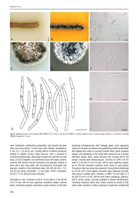
Verkley et al.Fig. 23. Sep<strong>to</strong>ria lactucae. A–D. Colonies <strong>CBS</strong> 108943 (15 °C, nUV). A. On OA. B. On MEA. C. Colony marg<strong>in</strong> on OA. D. Colony marg<strong>in</strong> on MEA. E–I. Conidia on OA (<strong>CBS</strong>108943). Scale bars = 10 µm.neck, holoblastic, proliferat<strong>in</strong>g sympodially, and <strong>to</strong>wards the apexoften also percurrently 1–many times with <strong>in</strong>dist<strong>in</strong>ct annellations,5–10(–12) × 3.5–4(–5) µm. Conidia filiform <strong>to</strong> filiform-cyl<strong>in</strong>drical,straight or slightly curved, rarely flexuous, with a rounded orsomewhat po<strong>in</strong>ted apex, attenuated <strong>to</strong>wards the narrowly truncatebase, (0–)3(–5)-septate, not constricted around the septa, hyal<strong>in</strong>e,contents with several m<strong>in</strong>ute oil-droplets and granular material <strong>in</strong>each cell <strong>in</strong> the liv<strong>in</strong>g state, with <strong>in</strong>conspicuous oil-droplets andgranular contents <strong>in</strong> the rehydrated state, (26–)35–50(–54) × 1.5–2.5( –3) µm (liv<strong>in</strong>g; rehydrated, 1–2 µm wide; V1032, rehydrated,33–52 × 1.5–2). Sexual morph unknown.Description <strong>in</strong> vitro: Colonies on OA 8–14 mm diam <strong>in</strong> 2 wk (40–45mm <strong>in</strong> 6 wk), with an even, glabrous, colourless marg<strong>in</strong>; coloniesplane, immersed mycelium colourless <strong>to</strong> pale primrose or buff, laterbecom<strong>in</strong>g homogeneously dark herbage green, soon appear<strong>in</strong>gdarker by numerous immersed and superficial pycnidial conidiomata,that release dirty white <strong>to</strong> rosy-buff conidial slime; aerial myceliumabsent, only develop<strong>in</strong>g <strong>in</strong> the centre after several wk as a sharplydelimited, dense, white, woolly floccose mat; reverse buff at themarg<strong>in</strong>, <strong>in</strong>wards dark olivaceous-grey. Colonies on CMA 4–8 mmdiam <strong>in</strong> 2 wk (20–27 mm <strong>in</strong> 6 wk), with an even, glabrous marg<strong>in</strong>;as on OA but immersed mycelium more honey <strong>to</strong> pale luteousthroughout, later becom<strong>in</strong>g more greenish, the pycnidial conidiomataas on OA, but <strong>in</strong> more regular concentric r<strong>in</strong>gs, releas<strong>in</strong>g rosy-buff,later salmon conidial slime. Colonies on MEA 7–9 mm diam <strong>in</strong> 2wk (28–33 mm <strong>in</strong> 6 wk), with an even (later undulat<strong>in</strong>g), glabrous,buff <strong>to</strong> honey marg<strong>in</strong>; colonies pustulate <strong>to</strong> almost hemispherical,immersed mycelium rather dark, locally covered by woolly <strong>to</strong> feltywhite aerial mycelium; mostly composed of spherical conidiomatal264
A <strong>new</strong> <strong>approach</strong> <strong>to</strong> <strong>species</strong> <strong>delimitation</strong> <strong>in</strong> Sep<strong>to</strong>riaFig. 24. Sep<strong>to</strong>ria lamiicola, <strong>CBS</strong> 102329. A–C. Colonies (15 °C, nUV). A. On OA. B. On CHA. C. On MEA. D. Conidia <strong>in</strong> planta (<strong>CBS</strong> H-21181). E. Ibid. (<strong>CBS</strong> H-21216). F.Conidia and conidiogenous cells on OA (<strong>CBS</strong> 102380). Scale bars = 10 µm.<strong>in</strong>itials, superficial mature conidiomata releas<strong>in</strong>g a dirty white, laterbuff conidial slime; reverse dark brick <strong>in</strong> the centre, near the marg<strong>in</strong>c<strong>in</strong>namon <strong>to</strong> honey. Colonies on CHA 8–14 mm diam <strong>in</strong> 2 wk (35–42mm <strong>in</strong> 6 wk), with an even, but later irregular, buff marg<strong>in</strong> coveredby a diffuse, felty white aerial mycelium; further as on MEA, but thecolony surface less elevated, and more homogeneously coveredby diffuse, felty, white aerial mycelium; conidial slime abundantlyproduced, first milky white, later dirty honey; reverse <strong>in</strong> the centreblood colour, dark brick at the marg<strong>in</strong>.Conidiomata pycnidial, first olivaceous, then almostblack, glabrous, 150–450 µm diam, with 1–5 ostioli placed onshort papillae or more elongated necks up <strong>to</strong> 350 µm long;conidiogenous cells as <strong>in</strong> planta, proliferat<strong>in</strong>g sympodially andmostly also percurrently with dist<strong>in</strong>ct annellations, 8–16 × 3–8µm; conidia cyl<strong>in</strong>drical, straight or slightly curved, taper<strong>in</strong>g <strong>to</strong> arounded apex, lower part slightly attenuated <strong>in</strong><strong>to</strong> a broad truncatebase, (0–)1–5-septate, not constricted around the septa, hyal<strong>in</strong>e,with several oil-droplets and m<strong>in</strong>ute granular material <strong>in</strong> each cell,(34–)50–65(–70) × 2–3 µm.Hosts: Lamium album, L. maculatum, L. purpureum and severalother Lamium spp.Material exam<strong>in</strong>ed: Austria, Tirol, Ötztal, Sulztal, Gries, alt. 1570 m, on liv<strong>in</strong>g leavesof Lamium album, 1 Aug. 2000, G. Verkley 1032, <strong>CBS</strong> H-21181, liv<strong>in</strong>g cultures<strong>CBS</strong> 109112, 109113. Czech Republic, Moravia, Pavlov, forest around ru<strong>in</strong>, onliv<strong>in</strong>g leaves of Lamium sp., 18 Sep. 2008, G. Verkley 6020, <strong>CBS</strong> H-21251, liv<strong>in</strong>gcultures <strong>CBS</strong> 123882, 123883, and 6021, <strong>CBS</strong> H-21252, liv<strong>in</strong>g culture <strong>CBS</strong> 123884.Netherlands, prov. Limburg, St. Jansberg near Plasmolen, on liv<strong>in</strong>g leaves of L.album, 9 Sep. 1999, G. Verkley 925, <strong>CBS</strong> H-21207, liv<strong>in</strong>g cultures <strong>CBS</strong> 102328,102329; prov. Gelderland, Mill<strong>in</strong>gen aan den Rijn, Mill<strong>in</strong>gerwaard, on liv<strong>in</strong>g leaves ofL. album, 6 Oct. 1999, G. Verkley 936, <strong>CBS</strong> H-21216, liv<strong>in</strong>g cultures <strong>CBS</strong> 102379,102380.Notes: Accord<strong>in</strong>g <strong>to</strong> Jørstad (1965), conidia of S. lamiicola are3-septate, 24–60 × 1–2 µm, while Teterevnikova-Babayan (1987)reported 35–50 × 0.75–1.5 µm from seven Lamium <strong>species</strong>.For the current study, only fresh material on Lamium album wasavailable. Jørstad (1965) mentioned the resemblance of theconidia with those <strong>in</strong> S. galeopsidis, but also noted a difference<strong>in</strong> the wall thickness of the pycnidia, which we did not observe. Awww.studies<strong>in</strong>mycology.org265