Verkley et al.<strong>to</strong> 60 µm diam, lack<strong>in</strong>g dist<strong>in</strong>ctly differentiated cells; conidiomatalwall composed of textura angularis without dist<strong>in</strong>ctly differentiatedlayers, mostly 15–20 µm thick, the outer cells with brown,somewhat thickened walls and 4.5–10 µm diam, the <strong>in</strong>ner cellshyal<strong>in</strong>e and th<strong>in</strong>-walled and of comparable diam. Conidiogenouscells hyal<strong>in</strong>e, discrete or <strong>in</strong>tegrated <strong>in</strong> short, 1–2-septateconidiophores, cyl<strong>in</strong>drical, or ampuliform with a relatively shortneck, hyal<strong>in</strong>e, holoblastic, proliferat<strong>in</strong>g sympodially, and sometimesalso percurrently with <strong>in</strong>dist<strong>in</strong>ct annellations, 6.5–10(–12.5) × 2.5–4.5 µm. Conidia cyl<strong>in</strong>drical, weakly <strong>to</strong> strongly curved, or flexuous,gradually attenuated <strong>to</strong> a rounded apex, gradually or more abruptlyattenuated <strong>in</strong><strong>to</strong> a broadly truncate base, (0–)2–5(–6)-septate, no<strong>to</strong>r <strong>in</strong>dist<strong>in</strong>ctly constricted around the septa, hyal<strong>in</strong>e, contents withseveral small guttules and numerous granules <strong>in</strong> each cell <strong>in</strong> theliv<strong>in</strong>g state, oil-droplets rarely merged <strong>in</strong><strong>to</strong> larger guttules <strong>in</strong> therehydrated state, (20–)40–65 × 2–2.5(–3) µm (rehydrated). Sexualmorph unknown.Description <strong>in</strong> vitro: Colonies on OA 7–10 mm diam <strong>in</strong> 2 wk (22–26 mm <strong>in</strong> 6 wk), with an even, somewhat undulat<strong>in</strong>g, glabrous,colourless marg<strong>in</strong>; colonies spread<strong>in</strong>g, the surface plane, immersedmycelium pale luteous or buff, with scattered immersed andsuperficial pycnidial conidiomata, which are first dark olivaceous,then almost black, glabrous, 150–450 µm diam, with a s<strong>in</strong>gle orseveral (up <strong>to</strong> 5!) ostioli placed on short papillae or more elongatednecks (up <strong>to</strong> 350 µm), that release buff <strong>to</strong> rosy-buff later salmonconidial slime; aerial mycelium diffuse, woolly-floccose, white;reverse honey, but isabell<strong>in</strong>e <strong>to</strong> hazel <strong>in</strong> the centre. Colonies onCMA 6–8 mm diam <strong>in</strong> 2 wk (18–23 mm <strong>in</strong> 6 wk), with an even,glabrous marg<strong>in</strong>; as on OA but immersed mycelium with a greenishhaze; aerial mycelium higher and reverse darker, later hazel witholivaceous and yellow t<strong>in</strong>ges; conidiomata similar as on OA.Colonies on MEA 7–9 mm diam <strong>in</strong> 2 wk (18–21 mm <strong>in</strong> 6 wk), withan even or somewhat undulat<strong>in</strong>g, glabrous, buff <strong>to</strong> honey marg<strong>in</strong>;colonies pustulate <strong>to</strong> almost hemispherical, immersed myceliumrather dark, near the marg<strong>in</strong> covered by woolly <strong>to</strong> felty white aerialmycelium; mostly composed of spherical conidiomatal <strong>in</strong>itials,superficial mature conidiomata releas<strong>in</strong>g rosy-buff <strong>to</strong> salmon, laterhoney conidial slime; reverse dark brick <strong>in</strong> the centre, near themarg<strong>in</strong> c<strong>in</strong>namon <strong>to</strong> honey. Colonies on CHA 7–14 mm diam <strong>in</strong> 2wk (20–28 mm <strong>in</strong> 6 wk), with an irregular, buff marg<strong>in</strong> covered by adiffuse, felty white, later grey aerial mycelium; further as on MEA,but the colony surface less elevated and especially near the marg<strong>in</strong>with greyish felty <strong>to</strong> tufty aerial mycelium; conidial slime abundantlyproduced, first rosy-buff, later salmon <strong>to</strong> ochreous; reverse <strong>in</strong> thecentre blood colour, dark brick <strong>to</strong> c<strong>in</strong>namon at the marg<strong>in</strong>.Conidiomata on OA see above. Conidia as <strong>in</strong> planta, mostly(0–)3–5(–6)-septate, 44–63(–70) × 2.5–3 µm.Hosts: Senecio fluviatilis and S. nemorensis.Material exam<strong>in</strong>ed: Belgium, Château de Namur, on leaves of Senecio sarracenica,1829, A. Bellynck, isotype BR-MYCO 155500-09. Netherlands, Prov. Gelderland,Mill<strong>in</strong>gen a/d Rijn, Mill<strong>in</strong>gerwaard, on liv<strong>in</strong>g leaves of S. fluviatilis, 6 Oct. 1999, G.Verkley 939, epitype designated here <strong>CBS</strong> H-21219 “MBT175358”, liv<strong>in</strong>g culturesex-epitype <strong>CBS</strong> 102366, 102381.Notes: The first Sep<strong>to</strong>ria that was described on the genus Seneciowas S. senecionis. The type host is Senecio sarracenica (=Senecio fluviatilis), and <strong>in</strong> later literature it has also been reportedfrom several other <strong>species</strong> of Senecio (Radulescu et al. 1973).Accord<strong>in</strong>g <strong>to</strong> the diagnosis by Westendorp, the conidia are 40 ×1.5 µm and 3–4-septate. Vanev et al. (1997) described the conidiaof S. senecionis as 2–6-septate, 29–68 × 2–2.5 µm, Radulescuet al. (1973) as 3–4-septate, 33–57 × 1.2–2 µm. By exam<strong>in</strong><strong>in</strong>gthe type specimen from BR it is here confirmed that conidia are <strong>in</strong>fact wider than described by Westendorp. It conta<strong>in</strong>s a s<strong>in</strong>gle leafwith a few lesions, and conidia observed are 30–55 × 1.5–2.5 µm,and mostly 3–5-septate. The fresh material that was collected <strong>in</strong>the Netherlands from the same host <strong>species</strong>, Senecio fluviatilis,and from which <strong>CBS</strong> 102366 and 102381 were isolated, is <strong>in</strong>sufficient agreement with the type and is therefore designated hereas epitype of S. senecionis. Differences with Sep<strong>to</strong>ria putrida arediscussed under that <strong>species</strong>.Sep<strong>to</strong>ria sii Roberge ex Desm., Pl. crypt. Fr., Fasc. 44, no2185; Annls Sci. Nat., sér. 3, Bot. 20: 92. 1853. Fig. 37.Description <strong>in</strong> planta: Symp<strong>to</strong>ms leaf spots, yellow <strong>to</strong> brown, <strong>in</strong>itiallyvaguely delimited but later well-delimited by ve<strong>in</strong>lets, scattered,later often confluent over large areas, visible on both sides of theleaf. Conidiomata pycnidial, epiphyllous, rarely also hypophyllous,s<strong>in</strong>gle, scattered or <strong>in</strong> small clusters, globose <strong>to</strong> subglobose,immersed, (60–)80–110 µm diam; ostiolum circular, central, 12.5–25(–35) µm wide, surround<strong>in</strong>g cells concolorous; conidiomatal wallcomposed of textura angularis 5–10 µm thick, with an outer layer ofcells 3–4.5 µm diam with brown, thickened walls, and an <strong>in</strong>ner layerof hyal<strong>in</strong>e and th<strong>in</strong>-walled cells, 2.5–4 µm diam. Conidiogenouscells hyal<strong>in</strong>e, broadly or elongated ampulliform, normally witha dist<strong>in</strong>ct neck, hyal<strong>in</strong>e, holoblastic, proliferat<strong>in</strong>g percurrently,annellations <strong>in</strong>dist<strong>in</strong>ct, 5–8.5 × 3–5 µm. Conidia cyl<strong>in</strong>drical, straight,curved, or flexuous, gradually attenuated <strong>to</strong> a relatively broadlyrounded apex, more or less abruptly attenuated <strong>in</strong><strong>to</strong> a truncatebase, 1–3(–4)-septate, slightly <strong>to</strong> dist<strong>in</strong>ctly constricted around thesepta <strong>in</strong> the fresh, fully hydrated state, hyal<strong>in</strong>e, conta<strong>in</strong><strong>in</strong>g one <strong>to</strong>several relatively large oil-droplets <strong>in</strong> each cell, <strong>in</strong> the rehydratedstate with irregular oil-masses (20–)29–35(–42) × 2–2.5(–3) µm(liv<strong>in</strong>g; rehydrated, 1.5–2 µm wide). Sexual morph unknown.Description <strong>in</strong> vitro: Colonies on OA 4–9 mm diam <strong>in</strong> 2 wk [(15–)19–23 mm <strong>in</strong> 6 wk], with an even, glabrous, colourless marg<strong>in</strong>;colonies rema<strong>in</strong><strong>in</strong>g almost plane, immersed mycelium olivaceousblack,locally however peach is dom<strong>in</strong>ant, which becomes scarletafter several wk; aerial mycelium mostly well-developed, woollyfloccose,white; scattered, mostly immersed pycnidial <strong>to</strong> stromaticconidiomata develop<strong>in</strong>g <strong>in</strong> the centre, releas<strong>in</strong>g droplets ofmilky white <strong>to</strong> rosy-buff conidial slime; reverse dark slate blue <strong>to</strong>olivaceous-black, and locally peach, the pigment not diffus<strong>in</strong>g <strong>in</strong><strong>to</strong>the medium. Colonies on CMA up <strong>to</strong> 1.5 mm diam <strong>in</strong> 2 wk [7–10(–25) mm <strong>in</strong> 6 wk], as on OA, but peach pigment diffus<strong>in</strong>g <strong>in</strong><strong>to</strong> themedium, while the colony itself is predom<strong>in</strong>antly olivaceous-black.Colonies frequently develop faster grow<strong>in</strong>g sec<strong>to</strong>rs that first arebuff and sporulate directly from the mycelium, later become paleluteous with a dist<strong>in</strong>ct scarlet pigmentation and form<strong>in</strong>g numerousmostly superficial pycnidia. Colonies on MEA 3–6 mm diam <strong>in</strong>2 wk [12–14(–26) mm <strong>in</strong> 6 wk], the marg<strong>in</strong> ruffled, olivaceousblack;colony concolorous, irregularly pustulate-worty, covered bydiffuse <strong>to</strong> dense felty white or greyish aerial mycelium; numerousconidiomatal <strong>in</strong>itials develop<strong>in</strong>g at the surface, mature onesreleas<strong>in</strong>g cirrhi of conidia that first are milky white, later salmon,sometimes merg<strong>in</strong>g <strong>to</strong> form slimy masses cover<strong>in</strong>g areas of thecolony surface; the agar surround<strong>in</strong>g the colony slightly discolouredby diffus<strong>in</strong>g pigment(s). Colonies on CHA 5–6 mm diam <strong>in</strong> 2 wk[8–13(–15) mm <strong>in</strong> 6 wk], as on MEA; some parts of the colonies286
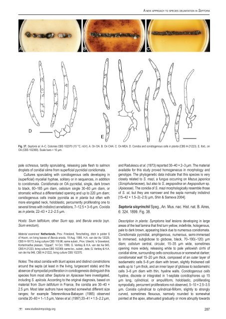
A <strong>new</strong> <strong>approach</strong> <strong>to</strong> <strong>species</strong> <strong>delimitation</strong> <strong>in</strong> Sep<strong>to</strong>riaFig. 37. Sep<strong>to</strong>ria sii. A–C. Colonies <strong>CBS</strong> 102370 (15 °C, nUV). A. On OA. B. On CHA. C. On MEA. D. Conidia and conidiogenous cells <strong>in</strong> planta (<strong>CBS</strong> H-21223). E. Ibid., onOA (<strong>CBS</strong> 102369). Scale bars = 10 µm.pale ochreous, tardily sporulat<strong>in</strong>g, releas<strong>in</strong>g pale flesh <strong>to</strong> salmondroplets of conidial slime from superficial pycnidial conidiomata.Cultures sporulat<strong>in</strong>g with conidiogenous cells develop<strong>in</strong>g <strong>in</strong>(superficial) mycelial hyphae, solitary or <strong>in</strong> sequences, <strong>in</strong> addition<strong>to</strong> conidiomata. Conidiomata on OA pycnidial, s<strong>in</strong>gle, dark brown<strong>to</strong> black, 80–185 µm diam, ostiolum s<strong>in</strong>gle 30–60 µm diam, orstromatic without a differentiated open<strong>in</strong>g and up <strong>to</strong> 220 µm diam;conidiogenous cells <strong>in</strong>side pycnidia as <strong>in</strong> planta but often withmore elongated neck, holoblastic, percurrently proliferat<strong>in</strong>g one <strong>to</strong>several times with <strong>in</strong>dist<strong>in</strong>ct annellations, 7–12.5 × 3–6 µm. Conidiaas <strong>in</strong> planta, 22–43 × 2.2–2.5 µm.Hosts: Sium latifolium, other Sium spp. and Berula erecta (syn.Sium erectum).Material exam<strong>in</strong>ed: Netherlands, Prov. Friesland, Terschell<strong>in</strong>g, ditch <strong>in</strong> polder Sof Hoorn, on liv<strong>in</strong>g leaves of Berula erecta, 19 Aug. 1995, H.A. van der Aa 12029,<strong>CBS</strong> H-18173, liv<strong>in</strong>g culture <strong>CBS</strong> 118.96; same substr., Prov. Utrecht, ‘s Graveland,Kortenhoefse plassen, “Oppad”, 14 Oct. 1999, G. Verkley & H.A. van der Aa 945,<strong>CBS</strong> H-21223, liv<strong>in</strong>g culture <strong>CBS</strong> 102369; same loc., substr., date, G. Verkley & H.A.van der Aa 946, <strong>CBS</strong> H-21222, liv<strong>in</strong>g culture <strong>CBS</strong> 102370.Notes: The s<strong>to</strong>ut conidia with blunt apices and dist<strong>in</strong>ct constrictionsaround the septa (at least <strong>in</strong> the liv<strong>in</strong>g, turgescent state) and theabsence of sympodial proliferation <strong>in</strong> conidiogenesis dist<strong>in</strong>guish this<strong>species</strong> from most other Sep<strong>to</strong>ria on Apiaceae here <strong>in</strong>vestigated,<strong>in</strong>clud<strong>in</strong>g S. apiicola. Accord<strong>in</strong>g <strong>to</strong> the orig<strong>in</strong>al diagnosis, based onmaterial from Sium latifolium <strong>in</strong> France, the conidia are 30–40 ×2.5 µm. Most later authors have reported somewhat different sizeranges; for example Teterevnikova-Babayan (1985) observedconidia 20–60 × 1–1.5 µm, Vanev et al. (1997) 20–41 × 1.5–2.2 µm,and Radulescu et al. (1973) reported 30–40 × 2–3 µm. The materialavailable for this study proved homogeneous <strong>in</strong> morphology andgenotype. The phylogenetic data <strong>in</strong>dicate that this <strong>species</strong> is veryclosely related <strong>to</strong> S. mazi, a fungus occurr<strong>in</strong>g on Mazus japonica(Scrophulariaceae), but also <strong>to</strong> S. aegopod<strong>in</strong>a on Aegopodium sp.(Apiaceae). The conidia of S. mazi morphologically resemble thoseof S. sii, but they are narrower and the septa normally <strong>in</strong>dist<strong>in</strong>ct[15–42 × 1.5–2(–2.5) µm, Sh<strong>in</strong> & Sameva 2004].Sep<strong>to</strong>ria sisyr<strong>in</strong>chii Speg., An. Mus. nac. Hist. nat. B. Aires,6: 324. 1899. Fig. 38.Description <strong>in</strong> planta: Symp<strong>to</strong>ms leaf lesions develop<strong>in</strong>g <strong>in</strong> largeareas of the leaf lam<strong>in</strong>a that first turn yellow, <strong>in</strong>def<strong>in</strong>ite, hologenous,pale <strong>to</strong> dark brown, appear<strong>in</strong>g black due <strong>to</strong> numerous conidiomata.Conidiomata pycnidial, amphigenous, numerous, semi-immersed<strong>to</strong> immersed, subglobose <strong>to</strong> globose, black, 70–100(–120) µmdiam; ostiolum central, circular, 15–35 µm wide, sometimesopen<strong>in</strong>g more widely, releas<strong>in</strong>g white <strong>to</strong> pale yellowish cirrhi ofconidial slime, surround<strong>in</strong>g cells concolourous or somewhat darker;conidiomatal wall 15–20 µm thick, composed of an outer layer ofisodiametric cells 5–8 µm diam with brown, slightly thickened cellwalls up <strong>to</strong> 1 μm thick, and an <strong>in</strong>ner layer of globose <strong>to</strong> isodiametriccells 3–6 μm diam with th<strong>in</strong>, hyal<strong>in</strong>e walls. Conidiogenous cellshyal<strong>in</strong>e, discrete or <strong>in</strong>tegrated <strong>in</strong> 1-septate conidiophores up 15µm long, cyl<strong>in</strong>drical, or ampulliform, holoblastic, proliferat<strong>in</strong>gsympodially, percurrent proliferations not observed, 5–10 × 2.5–3.5µm. Conidia cyl<strong>in</strong>drical <strong>to</strong> cyl<strong>in</strong>drical-filiform, slightly <strong>to</strong> stronglycurved, sometimes flexuous, narrowly rounded <strong>to</strong> somewhatpo<strong>in</strong>ted at the apex, attenuated gradually or more abruptly <strong>to</strong>wardswww.studies<strong>in</strong>mycology.org287


