PDF file (text) - Cryptogamic Botany Company
PDF file (text) - Cryptogamic Botany Company
PDF file (text) - Cryptogamic Botany Company
Create successful ePaper yourself
Turn your PDF publications into a flip-book with our unique Google optimized e-Paper software.
200<br />
THE MARINE ALGÆ OF NEW ENGLAND.<br />
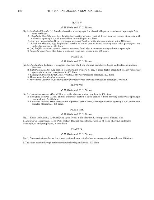
PLATE V.<br />
J. H. Blake and W. G. Farlow.<br />
Fig. 1. Leathcsia difformis, (L.) Aresch.; dissection showing a portion of cortical layer: a, a, unilocular sporangia; b, b,<br />
hairs. 400 diain.<br />
2. Chordaria flagelliformis, Ag.; longitudinal section of outer part of frond showing cortical filaments with<br />
unilocular sporangia, a, and a few cells of internal layer. 500 diam.<br />
3. Asperococcus echinatus, Grev.; transverse section of frond: a, unilocular sporangia; b, hairs. 150 diam.<br />
4. Stilophora rhizodes, Ag.; longitudinal section of outer part of frond showing sorus with paraphyses and<br />
unilocular sporangia. 400 diam.<br />
6. [sic] Ralfsia verrucosa, Aresch.; vertical section of frond with a sorus containing unilocular sporangia.<br />
6. Sphacelaria cirrhosa, (Roth) Ag.; a portion of frond with propagulum. 200 diam.<br />
PLATE VI.<br />
J. H. Blake and W. G. Farlow.<br />
Fig. 1. Chorda filum, L.; transverse section of portion of a frond showing paraphyses, b, and unilocular sporangia, a.<br />
200 diam.<br />
2. Stilophora rhizodes, Ag.; portion of sorus taken from Pl. V, Fig. 4, more highly magnified to show unilocular<br />
sporangia, a, a', and paraphyses, b. 600 diam.<br />
3. Ectocarpus littoralis, Lyngb., var. robustus, Farlow; plurilocular sporangia. 200 diam.<br />
4. The same with unilocular sporangia.<br />
5. Myrionenia Leclancheri, (Chauv.) Harv.; vertical section showing plurilocular sporangia.. 400 diam.<br />
PLATE VII.<br />
J. H. Blake and W. G. Farlow.<br />
Fig. 1. Castagnea virescens, (Carm.) Thuret; unilocular sporangium and hair, b. 400 diam.<br />
2. Castagnea Zosteræ, (Mohr.) Thuret; transverse section of outer portion of frond showing plurilocular sporangia,<br />
a, a', and hair, b. 400 diam.<br />
3. Elachistea fucicola, Fries; dissection of superficial part of frond, showing unilocular sporangia, a, a', and colored<br />
exserted filaments, b. 300 diam.<br />
PLATE VIII.<br />
J. H. Blake and W. G. Farlow.<br />
Fig. 1. Fucus vesiculosus, L.; fructifying tip of frond: a, air-bladder; b, conceptacles. Natural size.<br />
2. Laminaria longicruris, De la Pyl.; section through fructiferous portion of frond showing unilocular<br />
sporangia, a, and paraphyses, b. 400 diam.<br />
PLATE IX.<br />
J. H. Blake and W. G. Farlow.<br />
Fig. 1. Fucus vesiculosus, L.; section through a female conceptacle showing oospores and paraphyses. 200 diam.<br />
2. The same; section through male conceptacle showing antheridia. 200 diam.


