PDF file (text) - Cryptogamic Botany Company
PDF file (text) - Cryptogamic Botany Company
PDF file (text) - Cryptogamic Botany Company
Create successful ePaper yourself
Turn your PDF publications into a flip-book with our unique Google optimized e-Paper software.
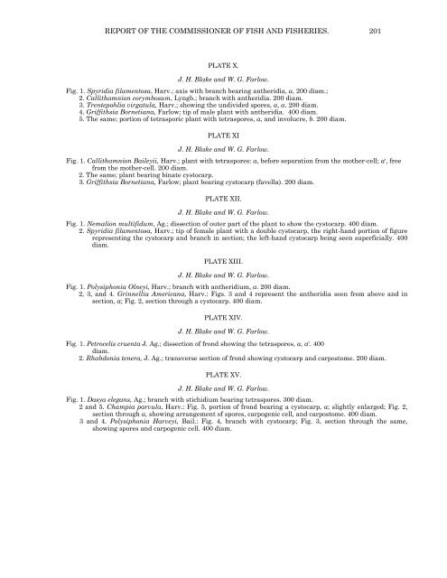
REPORT OF THE COMMISSIONER OF FISH AND FISHERIES. 201<br />
PLATE X.<br />
J. H. Blake and W. G. Farlow.<br />
Fig. 1. Spyridia filamentosa, Harv.; axis with branch bearing antheridia, a, 200 diam.;<br />
2. Callithamnion corymbosum, Lyngb.; branch with antheridia. 200 diam.<br />
3. Trentepohlia virgatula, Harv.; showing the undivided spores, a, a. 200 diam.<br />
4. Griffithsia Bornetiana, Farlow; tip of male plant with antheridia. 400 diam.<br />
5. The same; portion of tetrasporic plant with tetraspores, a, and involucre, b. 200 diam.<br />
PLATE XI<br />
J. H. Blake and W. G. Farlow.<br />
Fig. 1. Callithamnion Baileyii, Harv.; plant with tetraspores: a, before separation from the mother-cell; a', free<br />
from the mother-cell. 200 diam.<br />
2. The same; plant bearing binate cystocarp.<br />
3. Griffithsia Bornetiana, Farlow; plant bearing cystocarp (favella). 200 diam.<br />
PLATE XII.<br />
J. H. Blake and W. G. Farlow.<br />
Fig. 1. Nemalion multifidum, Ag.; dissection of outer part of the plant to show the cystocarp. 400 diam.<br />
2. Spyridia filamentosa, Harv.; tip of female plant with a double cystocarp, the right-hand portion of figure<br />
representing the cystocarp and branch in section; the left-hand cystocarp being seen superficially. 400<br />
diam.<br />
PLATE XIII.<br />
J. H. Blake and W. G. Farlow.<br />
Fig. 1. Polysiphonia Olneyi, Harv.; branch with antheridium, a. 200 diam.<br />
2, 3, and 4. Grinnellia Americana, Harv.: Figs. 3 and 4 represent the antheridia seen from above and in<br />
section, a; Fig. 2, section through a cystocarp. 400 diam.<br />
PLATE XIV.<br />
J. H. Blake and W. G. Farlow.<br />
Fig. 1. Petrocelis cruenta J. Ag.; dissection of frond showing the tetraspores, a, a'. 400<br />
diam.<br />
2. Rhabdonia tenera, J. Ag.; transverse section of frond showing cystocarp and carpostome. 200 diam.<br />
PLATE XV.<br />
J. H. Blake and W. G. Farlow.<br />
Fig. 1. Dasya elegans, Ag.; branch with stichidium bearing tetraspores. 300 diam.<br />
2 and 5. Champia parvula, Harv.: Fig. 5, portion of frond bearing a cystocarp, a; slightly enlarged; Fig. 2,<br />
section through a, showing arrangement of spores, carpogenic cell, and carpostome. 400 diam.<br />
3 and 4. Polysiphonia Harveyi, Bail.: Fig. 4, branch with cystocarp; Fig. 3, section through the same,<br />
showing spores and carpogenic cell. 400 diam.


