第117åæ¥æ¬è§£åå¦ä¼ç·ä¼ã»å ¨å½å¦è¡éä¼ è¬æ¼ããã°ã©ã ã»æé²é PDF ...
第117åæ¥æ¬è§£åå¦ä¼ç·ä¼ã»å ¨å½å¦è¡éä¼ è¬æ¼ããã°ã©ã ã»æé²é PDF ...
第117åæ¥æ¬è§£åå¦ä¼ç·ä¼ã»å ¨å½å¦è¡éä¼ è¬æ¼ããã°ã©ã ã»æé²é PDF ...
Create successful ePaper yourself
Turn your PDF publications into a flip-book with our unique Google optimized e-Paper software.
144<br />
117 <br />
P<br />
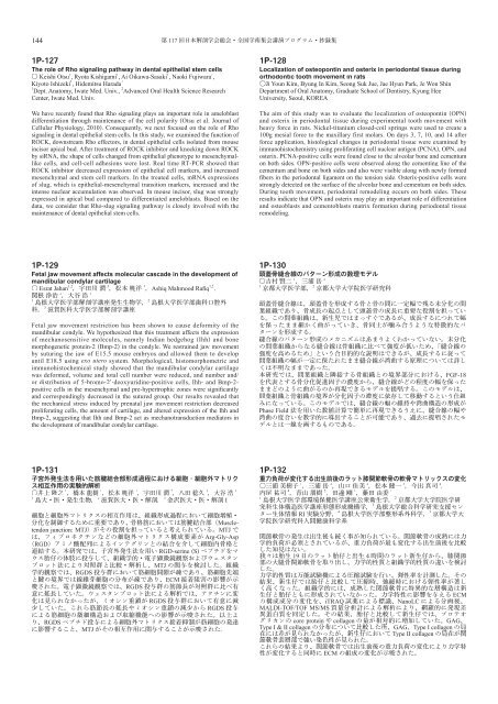
The role of Rho signaling pathway in dental epithelial stem cells<br />
Keishi Otsu 1 , Ryota Kishigami 1 , Ai OikawaSasaki 1 , Naoki Fujiwara 1 ,<br />
Kiyoto Ishizeki 1 , Hidemitsu Harada 1<br />
1<br />
Dept. Anatomy, Iwate Med. Univ., 2 Advanced Oral Health Science Research<br />
Center, Iwate Med. Univ.<br />
We have recently found that Rho signaling plays an important role in ameloblast<br />
differentiation through maintenance of the cell polarity Otsu et al. Journal of<br />
Cellular Physiology, 2010. Consequently, we next focused on the role of Rho<br />
signaling in dental epithelial stem cells. In this study, we examined the function of<br />
ROCK, downstream Rho effectors, in dental epithelial cells isolated from mouse<br />
incisor apical bud. After treatment of ROCK inhibitor and knocking down ROCK<br />
by siRNA, the shape of cells changed from epithelial phenotype to mesenchymal<br />
like cells, and cellcell adhesions were lost. Real time RTPCR showed that<br />
ROCK inhibitor decreased expression of epithelial cell markers, and increased<br />
mesenchymal and stem cell markers. In the treated cells, mRNA expressions<br />
of slug, which is epithelialmesenchymal transition markers, increased and the<br />
intense nuclear accumulation was observed. In mouse incisor, slug was strongly<br />
expressed in apical bud compared to differentiated ameloblasts. Based on the<br />
data, we consider that Rhoslug signaling pathway is closely involved with the<br />
maintenance of dental epithelial stem cells.<br />
P<br />
Localization of osteopontin and osterix in periodontal tissue during<br />
orthodontic tooth movement in rats<br />
Ji Youn Kim, Byung In Kim, Seong Suk Jue, Jae Hyun Park, Je Won Shin<br />
Department of Oral Anatomy, Graduate School of Dentistry, Kyung Hee<br />
University, Seoul, KOREA<br />
The aim of this study was to evaluate the localization of osteopontin OPN<br />
and osterix in periodontal tissue during experimental tooth movement with<br />
heavy force in rats. Nickeltitanium closedcoil springs were used to create a<br />
100g mesial force to the maxillary first molars. On days 3, 7, 10, and 14 after<br />
force application, histological changes in periodontal tissue were examined by<br />
immunohistochemistry using proliferating cell nuclear antigen PCNA, OPN, and<br />
osterix. PCNApositive cells were found close to the alveolar bone and cementum<br />
on both sides. OPNpositive cells were observed along the cementing line of the<br />
cementum and bone on both sides and also were visible along with newly formed<br />
fibers in the periodontal ligament on the tension side. Osterixpositive cells were<br />
strongly detected on the surface of the alveolar bone and cementum on both sides.<br />
During tooth movement, periodontal remodeling occurs on both sides. These<br />
results indicate that OPN and osterix may play an important role of differentiation<br />
and osteoblasts and cementoblasts matrix formation during periodontal tissue<br />
remodeling.<br />
P<br />
Fetal jaw movement affects molecular cascade in the development of<br />
mandibular condylar cartilage<br />
Esrat Jahan 1,2 3 1 Ashiq Mahmood Rafiq 1,2 <br />
2 1<br />
1<br />
2 <br />
3 <br />
Fetal jaw movement restriction has been shown to cause deformity of the<br />
mandibular condyle. We hypothesized that this treatment affects the expression<br />
of mechanosensitive molecules, namely Indian hedgehog Ihh and bone<br />
morphogenetic protein2 Bmp2 in the condyle. We restrained jaw movement<br />
by suturing the jaw of E15.5 mouse embryos and allowed them to develop<br />
until E18.5 using exo utero system. Morphological, histomorphometric and<br />
immunohistochemical study showed that the mandibular condylar cartilage<br />
was deformed, volume and total cell number were reduced, and number and/<br />
or distribution of 5bromo2′deoxyuridinepositive cells, Ihh and Bmp2<br />
positive cells in the mesenchymal and prehypertrophic zones were significantly<br />
and correspondingly decreased in the sutured group. Our results revealed that<br />
the mechanical stress induced by prenatal jaw movement restriction decreased<br />
proliferating cells, the amount of cartilage, and altered expression of the Ihh and<br />
Bmp2, suggesting that Ihh and Bmp2 act as mechanotransduction mediators in<br />
the development of mandibular condylar cartilage.<br />
P<br />
<br />
1 2<br />
1<br />
2 <br />
<br />
<br />
<br />
<br />
<br />
<br />
<br />
<br />
<br />
<br />
FGF18<br />
<br />
<br />
<br />
<br />
Phase Field <br />
<br />
<br />
P<br />
<br />
<br />
1 1 1 2 3 1<br />
1<br />
2 3 I<br />
<br />
Muscle<br />
tendon junction: MTJMTJ <br />
ArgGlyAsp<br />
RGD<br />
RGDserine S <br />
<br />
MTJ <br />
RGDS <br />
ECM <br />
RGDS <br />
<br />
RGDS <br />
RGDS <br />
<br />
RGDS <br />
MTJ <br />
P<br />
<br />
1 2 2 3 4 <br />
4 5 1 1<br />
1<br />
2 <br />
3 <br />
RI 4 5 <br />
<br />
<br />
<br />
<br />
19 4 <br />
<br />
<br />
<br />
<br />
<br />
ECM<br />
iTRAQQ<br />
NanoLC <br />
MALDITOF/TOF MS/MS <br />
<br />
core protein collagen GAG<br />
Type I & II collagen GAGType I collagen <br />
Type II collagen <br />
<br />
<br />
ECM


