211 Tabela 20A - Grupo submetido ao tratamento com gás argônio seguido pelo tratamento com hexafluoreto <strong>de</strong> enxofre (ArSF 6 /70W) Grupo Saliva Rugosida<strong>de</strong> Ângulo <strong>de</strong> contato Ângulo <strong>de</strong> contato cel/mm 2 log (cel) (Ra-μm) após tratamento (º) 48 horas (º) ARSF 6 Ausente 2,03 92,91 51,80 957,69 2,98 ARSF 6 Ausente 1,11 110,21 44,44 987,18 2,99 ARSF 6 Ausente 2,99 90,22 52,18 3705,13 3,57 ARSF 6 Ausente 1,72 103,60 54,25 578,21 2,76 ARSF 6 Ausente 2,19 102,12 48,19 648,72 2,81 ARSF 6 Ausente 1,20 104,94 58,02 346,15 2,54 ARSF 6 Ausente 1,05 93,44 49,30 1467,95 3,17 ARSF 6 Ausente 2,00 110,32 46,26 679,49 2,83 ARSF 6 Ausente 2,03 112,53 69,54 875,64 2,94 ARSF 6 Presente 1,01 97,56 48,80 3307,69 3,52 ARSF 6 Presente 1,97 97,15 57,80 2164,10 3,34 ARSF 6 Presente 1,70 99,40 52,21 2033,33 3,31 ARSF 6 Presente 2,34 85,52 60,34 2371,79 3,38 ARSF 6 Presente 1,82 98,57 55,78 4098,72 3,61 ARSF 6 Presente 2,04 109,96 70,51 1282,05 3,11 ARSF 6 Presente 1,71 101,38 49,29 6216,67 3,79 ARSF 6 Presente 1,10 98,52 57,02 206,41 2,32 ARSF 6 Presente 2,28 104,66 72,14 1671,79 3,22
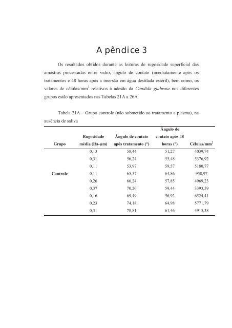
Apêndice 3 Os resultados obtidos durante as leituras <strong>de</strong> rugosida<strong>de</strong> superficial das amostras processadas entre vidro, ângulo <strong>de</strong> contato (imediatamente após os tratamentos e 48 horas após a imersão em água <strong>de</strong>stilada estéril), bem como, os valores <strong>de</strong> células/mm 2 relativos à a<strong>de</strong>são da Candida glabrata nos diferentes grupos estão apresentados nas Tabelas 21A a 26A. Tabela 21A – Grupo controle (não submetido ao tratamento a plasma), na ausência <strong>de</strong> saliva Ângulo <strong>de</strong> Grupo Rugosida<strong>de</strong> média (Ra-μm) Ângulo <strong>de</strong> contato após tratamento (°) contato após 48 horas (°) Células/mm 2 0,13 58,44 51,27 4039,74 0,31 56,24 55,48 5376,92 0,11 53,97 59,57 5180,77 Controle 0,11 65,57 64,86 958,97 0,26 66,24 57,85 4969,23 0,37 70,20 59,44 3393,59 0,16 69,49 56,92 6524,41 0,23 74,18 64,98 5771,79 0,31 78,81 61,46 4915,38
- Page 1 and 2:
UNIVERSIDADE ESTADUAL PAULISTA “J
- Page 3 and 4:
iii Dados Curriculares Camila Andra
- Page 5 and 6:
v exemplos, de quem eu me orgulho m
- Page 7 and 8:
vii Agradecimentos Especiais Muito
- Page 9 and 10:
ix mais novo”! Se eu pudesse, tor
- Page 11 and 12:
xi Aos meus amigos de turma do curs
- Page 13 and 14:
xiii À Capes (Coordenação de Ape
- Page 15 and 16:
Sumário Resumo....................
- Page 17 and 18:
ii alterações significantes nos v
- Page 19:
ii adherence of Candida albicans, e
- Page 22 and 23:
21 como: não eliminação do micro
- Page 24 and 25:
23 mais hidrofóbica a superfície,
- Page 26:
25 2008), resultando em uma superf
- Page 30 and 31:
3 Capítulos 3.1 Capítulo 1 Adhere
- Page 32 and 33:
31 Introduction The inability of cu
- Page 34 and 35:
33 negatively charged surfaces 22 .
- Page 36 and 37:
35 Plasma Treatments After roughnes
- Page 38 and 39:
37 measurements were performed in a
- Page 40 and 41:
39 was prepared in acetone at 0.4 m
- Page 42 and 43:
41 forces, the development of the m
- Page 44 and 45:
43 adherence observed in the presen
- Page 46 and 47:
45 effect 27,30,42-44 or decreased
- Page 48 and 49:
47 3 Yildirim MS, Hasanreisoglu U,
- Page 50 and 51:
49 of Thai silk treated by SF 6 pla
- Page 52 and 53:
51 33 Taylor R, Maryan C, Verran J.
- Page 54 and 55:
53 47 Waters MGJ, Williams DW, Jagg
- Page 56 and 57:
55 Table 2 - Means and standard dev
- Page 58 and 59:
Figure 2 57
- Page 60 and 61:
3.2 Capítulo 2 Evaluation of funga
- Page 62 and 63:
61 adhering to denture surfaces [1]
- Page 64 and 65:
63 mimicking the tissue-fitting sur
- Page 66 and 67:
65 spectroscopy analysis (XPS), car
- Page 68 and 69:
67 suspension was added to each wel
- Page 70 and 71:
69 Initial adherence of Candida alb
- Page 72 and 73:
71 increase probably occurred by me
- Page 74 and 75:
73 to the adsorption of salivary pr
- Page 76 and 77:
75 3) For all groups evaluated, no
- Page 78 and 79:
77 12. Pereira-Cenci T, Cury AADB,
- Page 80 and 81:
79 25. Ramage G, Tomsett K, Wickes
- Page 82 and 83:
81 38. Nikawa H, Hayashi S, Nikawa
- Page 84 and 85:
83 Tables Table 1 - Means and stand
- Page 86 and 87:
85 Figures Figure 1
- Page 88 and 89:
87 Figure Legends Figure 1 - XPS an
- Page 90 and 91:
89 Abstract Candida adhesion to pol
- Page 92 and 93:
91 to acrylic surfaces of Candida g
- Page 94 and 95:
93 minutes. The denture base acryli
- Page 96 and 97:
95 average was calculated. Specimen
- Page 98 and 99:
97 Statistical Analysis Differences
- Page 100 and 101:
99 the C. glabrata adhesion. The me
- Page 102 and 103:
101 One other important observation
- Page 104 and 105:
103 6. Minagi S, Miyake Y, Inagaki
- Page 106 and 107:
105 19. Nikawa H, Hayashi S, Nikawa
- Page 108 and 109:
107 33. Polukoshko KM, Brudvik JS,
- Page 110 and 111:
109 Tables Table 1 - Means and stan
- Page 112 and 113:
111 5.0 Log (cel/mm2) 4.0 3.0 2.0 *
- Page 114 and 115:
3.4 Capítulo 4 Effect of different
- Page 116 and 117:
115 Introduction The presence of Ca
- Page 118 and 119:
117 Italy) measuring 13.8 X 2 mm we
- Page 120 and 121:
119 4 - 6 ºC during the experiment
- Page 122 and 123:
121 Candida adhesion to dentures, a
- Page 124 and 125:
123 absorbance values obtained afte
- Page 126 and 127:
125 1. Silva WJ, Seneviratne J, Par
- Page 128 and 129:
127 14. Karaagaclioglu L, Can G, Yi
- Page 130 and 131:
129 28. Hahnel S, Rosentritt M, Han
- Page 132 and 133:
131 Tables Table 1 Table 1 - Median
- Page 134 and 135:
3.5 Capítulo 5 The effect of human
- Page 136 and 137:
135 Introduction An important step
- Page 138 and 139:
137 albicans adhesion to a denture
- Page 140 and 141:
139 For group G 3 , whole human uns
- Page 142 and 143:
141 each specimen, using a light mi
- Page 144 and 145:
143 was collected from one donor or
- Page 146 and 147:
145 In conclusion, this study focus
- Page 148 and 149:
147 11. Edgerton M, Scannapieco FA,
- Page 150 and 151:
149 24. Thein ZM, Samaranayake YH,
- Page 152 and 153:
151 37. Zamperini CA, Machado AL, V
- Page 154 and 155:
153 Figures Figure 1 *
- Page 156:
155 Figure Legends Figure 1. Mean a
- Page 159 and 160:
158 significantemente menores quand
- Page 161 and 162: 160 diminuindo os valores de ângul
- Page 163 and 164: 162 Nos estudos apresentados nos ca
- Page 165 and 166: 164 pela coloração cristal violet
- Page 167 and 168: 166 Diferentemente dos resultados o
- Page 170: 5 Conclusão Dentro das limitaçõe
- Page 173 and 174: 172 6. Bürgers R, Hahnel S, Reiche
- Page 175 and 176: 174 22. Hodak SK, Supasai T, Paosaw
- Page 177 and 178: 176 40. Nikawa H, Chen J, Hamada T,
- Page 179 and 180: 178 56. Pusateri CR, Monaco EA, Edg
- Page 181 and 182: 180 73. Tari BF, Nalbant D, Al DF,
- Page 184 and 185: 7 Anexos Anexo 1
- Page 186 and 187: Anexo 3
- Page 188 and 189: Anexo 5
- Page 190 and 191: Anexo 7
- Page 193 and 194: 8 Apêndice Apêndice 1 Os resultad
- Page 195 and 196: 194 Tabela 3A - Grupo submetido ao
- Page 197 and 198: 196 Tabela 5A - Grupo submetido ao
- Page 199 and 200: 198 Tabela 7A - Grupo submetido ao
- Page 201 and 202: 200 Tabela 9A - Grupo submetido ao
- Page 203 and 204: Apêndice 2 Os resultados obtidos d
- Page 205 and 206: 204 Tabela 13A - Grupo submetido ao
- Page 207 and 208: 206 Tabela 15A - Grupo submetido ao
- Page 209 and 210: 208 Tabela 17A - Grupo submetido ao
- Page 211: 210 Tabela 19A - Grupo submetido ao
- Page 215 and 216: 214 Tabela 24A - Grupo submetido ao
- Page 217 and 218: Apêndice 4 Os resultados obtidos d
- Page 219 and 220: 218 Tabela 30A - Grupo submetido ao
- Page 221 and 222: 220 Tabela 34A - Grupo submetido ao
- Page 223 and 224: Apêndice 5 Os resultados obtidos d
- Page 225 and 226: 224 Os resultados obtidos durante a
- Page 227: Autorizo a reprodução deste traba