Radiography in Modern Industry - Kodak
Radiography in Modern Industry - Kodak
Radiography in Modern Industry - Kodak

Create successful ePaper yourself
Turn your PDF publications into a flip-book with our unique Google optimized e-Paper software.
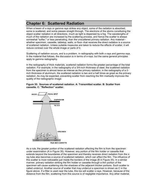
Chapter 6: Scattered RadiationWhen a beam of x-rays or gamma rays strikes any object, some of the radiation is absorbed,some is scattered, and some passes straight through. The electrons of the atoms constitut<strong>in</strong>g theobject scatter radiation <strong>in</strong> all directions, much as light is dispersed by a fog. The wavelengths ofmuch of the radiation are <strong>in</strong>creased by the scatter<strong>in</strong>g process, and hence the scatter is alwayssomewhat "softer," or less penetrat<strong>in</strong>g, than the unscattered primary radiation. Any material--whether specimen, cassette, tabletop, walls, or floor--that receives the direct radiation is a sourceof scattered radiation. Unless suitable measures are taken to reduce the effects of scatter, it willreduce contrast over the whole image or parts of it.Scatter<strong>in</strong>g of radiation occurs, and is a problem, <strong>in</strong> radiography with both x-rays and gamma rays.In the material that follows, the discussion is <strong>in</strong> terms of x-rays, but the same general pr<strong>in</strong>ciplesapply to gamma radiography.In the radiography of thick materials, scattered radiation forms the greater percentage of the totalradiation. For example, <strong>in</strong> the radiography of a 3/4-<strong>in</strong>ch thickness of steel, the scattered radiationfrom the specimen is almost twice as <strong>in</strong>tense as the primary radiation; <strong>in</strong> the radiography of a 2-<strong>in</strong>ch thickness of alum<strong>in</strong>um, the scattered radiation is two and a half times as great as the primaryradiation. As may be expected, prevent<strong>in</strong>g scatter from reach<strong>in</strong>g the film markedly improves thequality of the radiographic image.Figure 35: Sources of scattered radiation. A: Transmitted scatter. B: Scatter fromcassette. C: "Reflection" scatter.As a rule, the greater portion of the scattered radiation affect<strong>in</strong>g the film is from the specimenunder exam<strong>in</strong>ation (A <strong>in</strong> Figure 35). However, any portion of the film holder or cassette thatextends beyond the boundaries of the specimen and thereby receives direct radiation from the x-ray tube also becomes a source of scattered radiation, which can affect the film. The <strong>in</strong>fluence ofthis scatter is most noticeable just <strong>in</strong>side the borders of the image (B <strong>in</strong> Figure 35). In a similarmanner, primary radiation strik<strong>in</strong>g the film holder or cassette through a th<strong>in</strong> portion of thespecimen will cause scatter<strong>in</strong>g <strong>in</strong>to the shadows of the adjacent thicker portions. Such scatter iscalled undercut. Another source of scatter that may undercut a specimen is shown as C <strong>in</strong> thefigure above. If a filter is used near the tube, this too will scatter x-rays. However, because of thedistance from the film, scatter<strong>in</strong>g from this source is of negligible importance. Any other material,