Protocols - Hemorio
Protocols - Hemorio
Protocols - Hemorio

You also want an ePaper? Increase the reach of your titles
YUMPU automatically turns print PDFs into web optimized ePapers that Google loves.
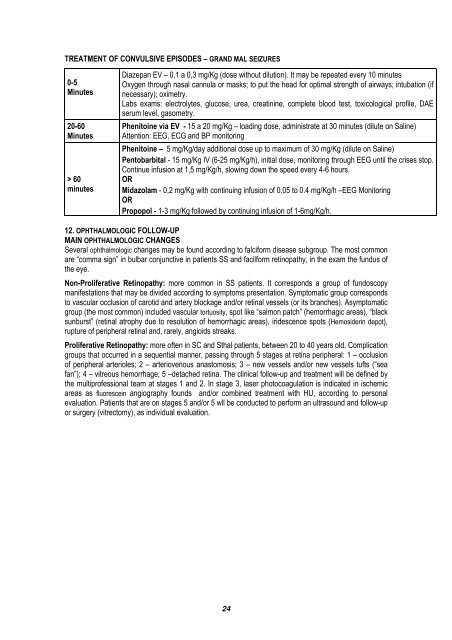
TREATMENT OF CONVULSIVE EPISODES – GRAND MAL SEIZURES<br />
0-5<br />
Minutes<br />
20-60<br />
Minutes<br />
> 60<br />
minutes<br />
Diazepan EV – 0,1 a 0,3 mg/Kg (dose without dilution). It may be repeated every 10 minutes<br />
Oxygen through nasal cannula or masks; to put the head for optimal strength of airways; intubation (if<br />
necessary); oximetry.<br />
Labs exams: electrolytes, glucose, urea, creatinine, complete blood test, toxicological profile, DAE<br />
serum level, gasometry.<br />
Phenitoine via EV - 15 a 20 mg/Kg – loading dose, administrate at 30 minutes (dilute on Saline)<br />
Attention: EEG, ECG and BP monitoring<br />
Phenitoine – 5 mg/Kg/day additional dose up to maximum of 30 mg/Kg (dilute on Saline)<br />
Pentobarbital - 15 mg/Kg IV (6-25 mg/Kg/h), initial dose, monitoring through EEG until the crises stop.<br />
Continue infusion at 1,5 mg/Kg/h, slowing down the speed every 4-6 hours.<br />
OR<br />
Midazolam - 0,2 mg/Kg with continuing infusion of 0.05 to 0.4 mg/Kg/h –EEG Monitoring<br />
OR<br />
Propopol - 1-3 mg/Kg followed by continuing infusion of 1-6mg/Kg/h.<br />
12. OPHTHALMOLOGIC FOLLOW-UP<br />
MAIN OPHTHALMOLOGIC CHANGES<br />
Several ophthalmologic changes may be found according to falciform disease subgroup. The most common<br />
are “comma sign” in bulbar conjunctive in patients SS and facilform retinopathy, in the exam the fundus of<br />
the eye.<br />
Non-Proliferative Retinopathy: more common in SS patients. It corresponds a group of fundoscopy<br />
manifestations that may be divided according to symptoms presentation. Symptomatic group corresponds<br />
to vascular occlusion of carotid and artery blockage and/or retinal vessels (or its branches). Asymptomatic<br />
group (the most common) included vascular tortuosity, spot like “salmon patch” (hemorrhagic areas), “black<br />
sunburst” (retinal atrophy due to resolution of hemorrhagic areas), iridescence spots (Hemosiderin depot),<br />
rupture of peripheral retinal and, rarely, angioids streaks.<br />
Proliferative Retinopathy: more often in SC and Sthal patients, between 20 to 40 years old. Complication<br />
groups that occurred in a sequential manner, passing through 5 stages at retina peripheral: 1 – occlusion<br />
of peripheral arterioles; 2 – arteriovenous anastomosis; 3 – new vessels and/or new vessels tufts (“sea<br />
fan”); 4 – vitreous hemorrhage; 5 –detached retina. The clinical follow-up and treatment will be defined by<br />
the multiprofessional team at stages 1 and 2. In stage 3, laser photocoagulation is indicated in ischemic<br />
areas as fluorescein angiography founds and/or combined treatment with HU, according to personal<br />
evaluation. Patients that are on stages 5 and/or 5 wll be conducted to perform an ultrasound and follow-up<br />
or surgery (vitrectomy), as individual evaluation.<br />
24