VOLUM OMAGIAL - Facultatea de Ştiinţe ale Naturii şi Ştiinţe Agricole
VOLUM OMAGIAL - Facultatea de Ştiinţe ale Naturii şi Ştiinţe Agricole
VOLUM OMAGIAL - Facultatea de Ştiinţe ale Naturii şi Ştiinţe Agricole

Create successful ePaper yourself
Turn your PDF publications into a flip-book with our unique Google optimized e-Paper software.
Simona Ghiţă et al. / Ovidius University Annals, Biology-Ecology Series 14: 127-137 (2010)<br />
very low (and responsible for the low increase in the<br />
average cell lengths in M3). In M3 (control) one can<br />
see a rather constant length of some cells during<br />
incubation (2,15±0,37) whereas in M2 there was a<br />
sud<strong>de</strong>n increase in cell length (6,46±1,54) to the time<br />
T2 (4 hours incubation) then there was a steady<br />
increase until T4 (8 hours incubation).<br />
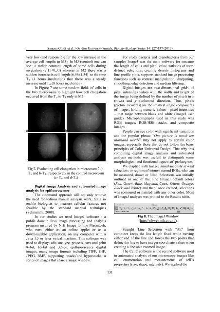
In Figure 7 are some random fields of cells in<br />
the two microcosms to highlight how cell elongation<br />
occurred from the T o to T4 only in M2.<br />
a b<br />
c d<br />
Fig 7. Evaluating cell elongation in microcosm 2 (a-<br />
To and b-T4) respectively in the control microcosm<br />
(c- To and d-T4)<br />
Digital Image Analysis and automated image<br />
analysis for epifluorescence<br />
The automated approach will not only remove<br />
the need for tedious manual analysis work, but also<br />
enable biologists to measure cellular features not<br />
feasible by the standard manual techniques<br />
(Selinummi, 2008).<br />
In our studies we used ImageJ software - a<br />
public domain Java image processing and analysis<br />
program inspired by NIH Image for the Macintosh,<br />
who runs, either as an online applet or as a<br />
downloadable application, on any computer with a<br />
Java 1.5 or later virtual machine. This software was<br />
used to display, edit, analyze, process, save and print<br />
8–bit, 16–bit and 32–bit epifluorescence digital<br />
images, many image formats including TIFF, GIF,<br />
JPEG, BMP, supporting ‘stacks’and hyperstacks, a<br />
series of images that share a single window.<br />
131<br />
For study bacteria and cyanobacteria from our<br />
samples ImageJ was the main software for measure<br />
the length of cells and pixel value statistics of user<strong>de</strong>fined<br />
selections, creating <strong>de</strong>nsity histograms and<br />
line profile plots, supports standard image processing<br />
functions such as contrast manipulation, sharpening,<br />
smoothing, edge <strong>de</strong>tection and median filtering.<br />
Digital images are two-dimensional grids of<br />
pixel intensities values with the width and height of<br />
the image being <strong>de</strong>fined by the number of pixels in x<br />
(rows) and y (columns) direction. Thus, pixels<br />
(picture elements) are the smallest single components<br />
of images, holding numeric values – pixel intensities<br />
– that range between black and white (ImageJ user<br />
gui<strong>de</strong>). Microphotographs used in this study was<br />
RGB images, RGB/HSB stacks, and composite<br />
images.<br />
People can see color with significant variations<br />
and the popular phrase “One picture is worth ten<br />
thousand words” may not apply to certain color<br />
images, especially those that do not follow the basic<br />
principles of Color Universal Design. That why this<br />
combining digital image analysis and automated<br />
analysis methods was usefull to distinguish some<br />
morphological and functional aspects of prokaryotes.<br />
We displied with ImageJ simultaneously several<br />
selections or regions of interest named ROIs, who can<br />
be measured, drawn or filled. Selections was initially<br />
outlined in one of the nine ImageJ <strong>de</strong>fault colors<br />
(Red, Green, Blue, Magenta, Cyan, Yellow, Orange,<br />
Black and White) and then, once created, selections<br />
was contoured or painted with any other color. Most<br />
of ImageJ analyses was printed to the Results table.<br />
Fig 8. The ImageJ Window<br />
(http://rsbweb.nih.gov/ij/).<br />
Straight Line Selection with “Alt” from<br />
computer keeps the line length fixed while moving<br />
either end of the line and forces the two points that<br />
<strong>de</strong>fine the line to have integer coordinate values when<br />
creating a line on a zoomed image.<br />
The CellC software is the second software used<br />
in automated analysis of our microscopy images like<br />
cell enumeration and measurements of cell’s<br />
properties (size, shape, intensity). We applied the