VOLUM OMAGIAL - Facultatea de Ştiinţe ale Naturii şi Ştiinţe Agricole
VOLUM OMAGIAL - Facultatea de Ştiinţe ale Naturii şi Ştiinţe Agricole
VOLUM OMAGIAL - Facultatea de Ştiinţe ale Naturii şi Ştiinţe Agricole

Create successful ePaper yourself
Turn your PDF publications into a flip-book with our unique Google optimized e-Paper software.
Utilization of epifluorescence microscopy…/ Ovidius University Annals, Biology-Ecology Series 14: 127-137 (2010)<br />
spring and heterotrophic and phototrophic bacteria<br />
from marine environment.<br />
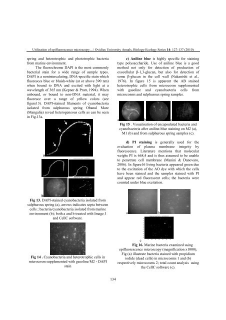
The fluorochrome DAPI is the most commonly<br />
bacterial stain for a wi<strong>de</strong> range of sample types.<br />
DAPI is a nonintercalating, DNA-specific stain which<br />
fluoresces blue or bluish-white (at or above 390 nm)<br />
when bound to DNA and excited with light at a<br />
wavelength of 365 nm (Kepner & Pratt, 1994). When<br />
unbound, or bound to non-DNA material, it may<br />
fluoresce over a range of yellow colors (see<br />
figure13). DAPI-stained filaments of cyanobacteria<br />
isolated from sulphurous spring Obanul Mare<br />
(Mangalia) reveal heterogeneous cells as can be seen<br />
in Fig.13a.<br />
a<br />
b<br />
Fig 13. DAPI-stained cyanobacteria isolated from<br />
sulphurous spring (a), arrows indicates septa between<br />
cells ; bacteria/cyanobacteria isolated from marine<br />
environment (b); both a and b treated with Image J<br />
and CellC software.<br />
Fig 14 . Cyanobacteria and heterotrophic cells in<br />
microcosm supplemented with gasoline/M2 - DAPI<br />
stain<br />
134<br />
c) Aniline blue is highly specific for staining<br />
type polysacchari<strong>de</strong>. Use of aniline blue is a good<br />
method not only for <strong>de</strong>tection of production of<br />
exocellular β-1,3-glucan, but also for <strong>de</strong>tection of<br />
some β-glucan in the cell wall (Nakanishi et al.,<br />
1976). In figure 15 is apparent the AB stained<br />
heterotrophic cells from microcosm supplemented<br />
with gasoline and cyanobacteria cells from<br />
microcosms and sulphurous spring samples.<br />
a b c<br />
Fig 15 . Visualisation of encapsulated bacteria and<br />
cyanobacteria after aniline-blue staining on M2 (a),<br />
M1 (b) and from sulphurous spring samples (c).<br />
d) PI staining is generally used for the<br />
evaluation of plasma membrane integrity by<br />
fluorescence. Literature mentions that molecular<br />
weighs PI is 668,4 and is thus assumed to be unable<br />
to penetrate cell membrane (Manini & Danovaro,<br />
2006). In figure16 living bacteria appeared green due<br />
to the excitation of the AO dye with which the cells<br />
have been stained and the samples stained with PI<br />
and appear red fluorescent cells; the bacteria were<br />
counted un<strong>de</strong>r blue excitation.<br />
a b<br />
c<br />
Fig 16. Marine bacteria examined using<br />
epifluorescence microscopy (magnification x1000),<br />
Fig (a) illustrate bacteria stained with propidium<br />
iodi<strong>de</strong> (<strong>de</strong>ad cells) in microcosms 1 and (b)<br />
respectively microcosms 2; total count analysis using<br />
the CellC software (c).