VOLUM OMAGIAL - Facultatea de Ştiinţe ale Naturii şi Ştiinţe Agricole
VOLUM OMAGIAL - Facultatea de Ştiinţe ale Naturii şi Ştiinţe Agricole
VOLUM OMAGIAL - Facultatea de Ştiinţe ale Naturii şi Ştiinţe Agricole

Create successful ePaper yourself
Turn your PDF publications into a flip-book with our unique Google optimized e-Paper software.
The clinical utility of aditional metho<strong>de</strong>s... / Ovidius University Annals, Biology-Ecology Series 14: 157-162 (2010)<br />
From all patients with histopathologic and<br />
clinical data that indicate malignancy, a number of 5<br />
patients (2 / 2, 100% associated with malignant<br />
mesothelioma, 1/4, 25% associated with hepatic<br />
carcinoma, ¼, 25% associated with breast carcinoma,<br />
1/3, 33.33% associated with lung carcinoma) were<br />
<strong>de</strong>ceased before drawing to the final study (a period<br />
of approximately three years from the accumulation<br />
of fluid in the peritoneal cavity), in all cases, the<br />
peritoneal fluid cytology recor<strong>de</strong>d the presence of<br />
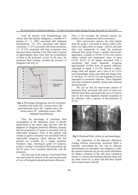
malignant cells (Fig. 4).<br />
Fig. 4. Percentage of prognostic survival of patients<br />
inclu<strong>de</strong>d in the study (OC- ovarian cancer, GIC -<br />
gastrointestinal cancer, HC - hepatic cancer, BC –<br />
breast cancer, PC - pulmonary cancer, MM -<br />
malignant mesothelioma)<br />
Thus, the percentage of peritoneal fluid<br />
accumulation in the abdominal cavity is directly<br />
proportional to the tumor stage and also with the<br />
diagnosis of malignant peritoneal effusions, meaning<br />
that the progression of cancer is associated with an<br />
unfavorable prognosis. None of the patients with<br />
ovarian or gastric carcinoma were associated with an<br />
unfavorable prognosis, which indicates that this<br />
patients are available for a longer treatment.<br />
Neoplasia stage, histological gra<strong>de</strong> of neoplasia,<br />
positive cytology of peritoneal fluid and patients age<br />
(61-70 years) were correlated statistically with the<br />
prognosis.<br />
The first step in the analysis of peritoneal fluid<br />
was represented by analysis of the macroscopic point<br />
of view of biological material (peritoneal effusions).<br />
160<br />
Thus, it was recor<strong>de</strong>d: the extracted amount, the<br />
product color, transparency and its consistency.<br />
After macroscopic analysis, the most liquids<br />
from the group I was found to shown yellow color<br />
(from very light yellow to orange - yellow), and most<br />
fluid were transparent. In stead, the peritoneal<br />
effusions from group II had a variable macroscopic<br />
appearance: a number of 29/40 (72.5%) were intense<br />
yellow colored and transparents, many of them<br />
(17/29, 42.5% of all liquids associated with a<br />
carcinoma) had tissue fragments occupying<br />
approximately 25-50% from all quantity effusions,<br />
suspen<strong>de</strong>d in liquid; 5 (12.5%) showed a yelloworange<br />
fluid and opaque, and a total of six (15%)<br />
were hemorrhagic (<strong>de</strong>ep red), fluid and opaque (Fig.<br />
5). Of these, 19 / 40 (47.5%) had fragments of tissue<br />
suspen<strong>de</strong>d in peritoneal effusions. These fragments<br />
were then inclu<strong>de</strong>d in paraffin, stained and examined<br />
microscopically.<br />
We can say that the macroscopic analysis of<br />
peritoneal fluid, associated with cases of cancer are<br />
different from those associated with cases of cirrhosis<br />
only by this tissue fragments foun<strong>de</strong>d suspen<strong>de</strong>d in<br />
the effusions, with a capacity of discrimination of<br />
47.5%.<br />
Fig. 5. Peritoneal fluid: yellow (a) and hemorrhagic<br />
The registration of the important differences<br />
existing between macroscopic peritoneal fluids is<br />
essential, representing the first way in effusions<br />
discrimination (in transudates and exudates), so that,<br />
the material subjected can provi<strong>de</strong> useful information<br />
for further evaluation of the cells from cytological<br />
smears. It is known that bleeding effusions (<strong>de</strong>ep red)<br />
are often caused by a cancer and that these liquids<br />
often contain cancer cells [9].