VOLUM OMAGIAL - Facultatea de Ştiinţe ale Naturii şi Ştiinţe Agricole
VOLUM OMAGIAL - Facultatea de Ştiinţe ale Naturii şi Ştiinţe Agricole
VOLUM OMAGIAL - Facultatea de Ştiinţe ale Naturii şi Ştiinţe Agricole

You also want an ePaper? Increase the reach of your titles
YUMPU automatically turns print PDFs into web optimized ePapers that Google loves.
Ana Maria Creţu et al. / Ovidius University Annals, Biology-Ecology Series 14: 157-162 (2010)<br />
However, comparing the results, after<br />
performing peritoneal fluid cytology, with those<br />
obtained by macroscopic evaluation, of the 40<br />
effusions associated with at least one malignancy,<br />
only 6 (15%) were foun<strong>de</strong>d to be red colored<br />
(hemorrhagic), emphasizing that it does not exist any<br />
relationship between peritoneal fluid containing<br />
cancer cells and fluid color.<br />
After conducting the Riwalta reactions [10], the<br />
81 peritoneal effusions were classified in: 29<br />
(35.80%) transudates peritoneal fluid (with low cell<br />
<strong>de</strong>nsity and low protein content, which is usually<br />
accumulated in benign conditions) and 43 (53.08%)<br />
exudate (effusions with high cell <strong>de</strong>nsity and high<br />
protein content, which is accumulated most in<br />
malignant conditions), and 9 (11.11%) mixed,<br />
intermediate peritoneal effusions. Thus, peritoneal<br />
fluids were classified into three groups: group I<br />
(transudates), group II (intermediate, mixed) and<br />
group III (exudates) (Table 2).<br />
Table 2. Distribution of cases after Riwalta reaction<br />
Lots Primary<br />
cancer<br />
Lot I<br />
(n=41)<br />
Lot II<br />
(n=40)<br />
Transudates<br />
(N=29)<br />
Mixed<br />
(N=9)<br />
Exudates<br />
(N=43)<br />
CB 21 3 17<br />
CH (4) 1 1 2<br />
CO (18) 3 0 15<br />
CGI (9) 1 2 6<br />
CM (4) 2 2 0<br />
CP (3) 1 1 1<br />
MP (2) 0 0 2<br />
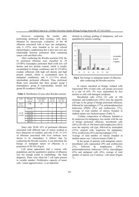
Since only 26/40, 65% of peritoneal effusions<br />
associated with different type of cancer resulted to<br />
have characters of exudates, and only 21/41, 51.21%<br />
of effusions associated with liver cirrhosis were<br />
shown to be transudates, it follows that, by<br />
conducting the Riwalta reaction, it can <strong>de</strong>termine the<br />
benign or malignant nature of effusions in a<br />
proportion of 58.10% (Fig.6).<br />
Cell smear appearance had a various cells<br />
populations and the quantitative analysis of effusions<br />
was not enough useful in establishing the final<br />
diagnosis. There were observed 7 cell types present<br />
in variable number. Proliferative capacity of tumor<br />
cells - tumor aggressiveness - is an important<br />
161<br />
element in cytology grading of malignancy, and was<br />
quantified by mitosis counting.<br />
Fig 6. The benign or malignant nature of effusions<br />
after conducting the Riwalta reaction<br />
In smears classified as benign, isolated cells<br />
represented 90% of total cells, cell groups recovered<br />
to a rate of 10%. 5% were represented by free<br />
nucleus or cells with damaged cytoplasm.<br />
Mesothelial cells (33%) (33 cells of 100<br />
elements) and lymphocytes (30%) were the majority<br />
cell type in the group of benign peritoneal effusions,<br />
followed by macrophages (17%), polymorphonuclear<br />
leukocytes (PMN) (9%) and erythrocytes (7%).<br />
Average of total number of mitosis foun<strong>de</strong>d in<br />
studied smears was 3mitosis/smears (Table 3).<br />
Cellular composition of effusions foun<strong>de</strong>d to<br />
be suspicious for malignancy was similar with the one<br />
of benign peritoneal effusions: mesothelial cells<br />
(28%) (28 cells of 100 items) and lymphocytes (26%)<br />
were the majority cell type, followed by neutrophils<br />
(13%), atypical cells, suspicious for malignancy<br />
(9%), erythrocytes (9%) and macrophages (7%).<br />
Average of total number of mitosis foun<strong>de</strong>d in<br />
studied smears was 7 mitosis/smears (Table 3).<br />
In the group of patients with malignant cancer,<br />
mesothelial cells represented 29% and erythrocytes<br />
21%, followed by lymphocytes (18%),<br />
polymorphonuclear leukocytes (16%), macrophages<br />
(4%) and malignant cells (4%), average of total<br />
number of mitosis was 9 mitoses / cell smear (Figure<br />
7).