VOLUM OMAGIAL - Facultatea de Ştiinţe ale Naturii şi Ştiinţe Agricole
VOLUM OMAGIAL - Facultatea de Ştiinţe ale Naturii şi Ştiinţe Agricole
VOLUM OMAGIAL - Facultatea de Ştiinţe ale Naturii şi Ştiinţe Agricole

You also want an ePaper? Increase the reach of your titles
YUMPU automatically turns print PDFs into web optimized ePapers that Google loves.
Simona Ghiţă et al. / Ovidius University Annals, Biology-Ecology Series 14: 127-137 (2010)<br />
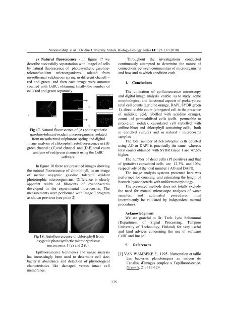
e) Natural fluorescence - In figure 17 we<br />
<strong>de</strong>scribe succesfully separatation with ImageJ of cells<br />
by natural fluorescence of photosynthetic gasolinetolerant/oxidant<br />
microorganisms isolated from<br />
mesothermal sulphurous spring in different chanell –<br />
red and green- and then each image were automat<br />
counted with CellC, obtaining finally the number of<br />
cells red and green separately.<br />
Fig 17. Natural fluorescence of (A) photosynthetic<br />
gasoline-tolerant/oxidant microorganisms isolated<br />
from mesothermal sulphurous spring and digital<br />
image analysis of chlorophyll autofluorescence in (B)<br />
green channel ; (C) red channel and (D-E) total count<br />
analysis of red/green channels using the CellC<br />
software.<br />
In figure 18 there are presented images showing<br />
the natural fluorescence of chlorophyll, as an image<br />
of marine oxygenic gasoline tolerant/ oxidant<br />
phototrophic microorganisms. Difference is clearly<br />
apparent width of filaments of cyanobacteria<br />
<strong>de</strong>veloped in the experimental microcosms. The<br />
measurements were performed with Image J program<br />
as shown previous (see point 2).<br />
a b<br />
Fig 18. Autofluorescence of chlorophyll from<br />
oxygenic photosynthetic microorganisms:<br />
microcosms 1 (a) and 2 (b).<br />
Epifluorescence techniques and image analysis<br />
has increasingly been used to <strong>de</strong>termine cell size,<br />
bacterial abundance and <strong>de</strong>tection of physiological<br />
characteristics like damaged versus intact cell<br />
membranes.<br />
135<br />
Throughout the investigations conducted<br />
continuously attempted to <strong>de</strong>termine the nature of<br />
connections between communities of microorganisms<br />
and how and to which condition each.<br />
4. Conclusions<br />
The utilization of epifluorescence microscopy<br />
and digital image analysis enable us to study some<br />
morphological and functional aspects of prokaryotes:<br />
total cell counts (acridine orange, DAPI, SYBR green<br />
1), direct viable count (elongated cell in the presence<br />
of nalidixic acid, labelled with acridine orange),<br />
count of permeabilised cells (cells permeable to<br />
propidium iodi<strong>de</strong>), capsulated cell (labelled with<br />
aniline blue) and chlorophyll containing cells, both<br />
in enriched cultures and in natural / microcosms<br />
samples.<br />
The total number of heterotrophic cells counted<br />
using AO or DAPI is practically the same whereas<br />
total counts obtained with SYBR Green 1 are 47,6%<br />
higher.<br />
The number of <strong>de</strong>ad cells (PI positive) and that<br />
of (putative) capsulated cells are 12.3% and 10%,<br />
respectively of the total number ( AO and DAPI).<br />
The image analysis systems presented here was<br />
performed for counting and estimating the length of<br />
bacteria/cyanobacteria with uniform morphology.<br />
The presented methods does not totally exclu<strong>de</strong><br />
the need for manual microscope analyses of water<br />
samples, and automated procedures must<br />
intermittently be validated by in<strong>de</strong>pen<strong>de</strong>nt manual<br />
procedures.<br />
Acknowledgment<br />
We are grateful to Dr. Tech. Jyrki Selinummi<br />
(Department of Signal Processing, Tampere<br />
University of Technology, Finland) for very useful<br />
and kind advices concerning the use of software<br />
CellC and ImageJ.<br />
5. References<br />
[1] VAN WAMBEKE F., 1995- Numeration et taille<br />
<strong>de</strong>s bacteries planctoniques au moyen <strong>de</strong><br />
l`analise d`images couplee a l`epifluorescence.<br />
Oceanis, 21: 113-124.