VOLUM OMAGIAL - Facultatea de Ştiinţe ale Naturii şi Ştiinţe Agricole
VOLUM OMAGIAL - Facultatea de Ştiinţe ale Naturii şi Ştiinţe Agricole
VOLUM OMAGIAL - Facultatea de Ştiinţe ale Naturii şi Ştiinţe Agricole

Create successful ePaper yourself
Turn your PDF publications into a flip-book with our unique Google optimized e-Paper software.
Simona Ghiţă et al. / Ovidius University Annals, Biology-Ecology Series 14: 127-137 (2010)<br />
cell). Means of each column and the unit of measure<br />
(pixels or micrometers) are presented in the end of<br />
the file.<br />
Image acquisition—Images for analysis were<br />
done with a Canon digital camera. Brightness and<br />
contrast were adjusted for the first image and kept<br />
unchanged throughout the image acquisition<br />
procedure. The images (1600 by 1200 pixels, 256<br />
dpi) were acquired at 50x magnification and stored as<br />
543-KB JPG files. Additional images acquired at<br />
100x magnification were used to verify that<br />
measurements of individual filaments/ bacteria were<br />
in<strong>de</strong>pen<strong>de</strong>nt of magnification<br />
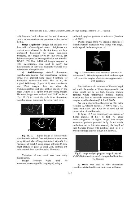
a) Acridin-orange stained filamentous<br />
cyanobacteria isolated from mesothermal sulfurous<br />
spring were analysed using Image J software for<br />
distinguish heterocystous cells. First of all, the<br />
original RGB image (Figure 10 A) were transformed<br />
into 32-bit images, then we adjust the<br />
brightness/contrast and also applied smooth or find<br />
edges (Figure 10 B) option from processing images.<br />
The same image were analysed with CellC software<br />
(Fig. 10 C) to count the cells from filamentous<br />
cyanobacteria or to measure the size of each cells.<br />
A B<br />
C<br />
Fig 10. A – digital image of heterocystous<br />
cyanobacteria isolated from sulphurous mesothermal<br />
spring Obanul Mare (Mangalia) stained with AO; B –<br />
find edges of panel A using ImageJ software; C- total<br />
count analysis of panel A using CellC software (48<br />
cells counted from cyanobacteria’s filaments).<br />
Validation of any count were done using<br />
manual count.<br />
ImageJ software were used for<br />
automated measuring cell’s length (µm), using a<br />
133<br />
calibrated eyepiece graticule as reference (Ar<strong>de</strong>lean<br />
et al, 2009).<br />
Digital images from AO staining filaments of<br />
cyanobacteria in microcosm were treated with ImageJ<br />
to distinguish the heterocystous cell.<br />
Fig 11. Cyanobacteria with heterocyst presence in<br />
microcosm 2; AO staining (arrow indicate heterocyst<br />
cell present in samples of microcosm supplemented<br />
with gasoline).<br />
To avoid uncertain estimates of filament length<br />
and width, the number of filaments presented in one<br />
image should not be too high. Extreme filament<br />
<strong>de</strong>nsities would undoubtedly increase filament<br />
overlap and lead to uncertain measurements unless<br />
samples are diluted (Almesjö & Rolff, 2007).<br />
We use a blue light epifluorescence filter set to<br />
visualize AO-stained bacteria (N-400FL type). AO<br />
stains both DNA and RNA so is used for the<br />
enumeration of total bacteria.<br />
In figure 12 A we present only an example of<br />
digital analysis of fig.7 b: first, we adjust<br />
contrast/brightness of digital image, then analyse<br />
measure of graticula presented in fig. 7b and set the<br />
calibration bar to <strong>de</strong>termine correctly the length of<br />
each bacteria treated with nalidixic acid. In B is<br />
presented image analysis using CellC software.<br />
A B<br />
Fig 12. Image analysis program Image J (A) and<br />
CellC (B) from microcosm 2, elongated cells at time<br />
T4 (8hours)<br />
b) DAPI were used to view filamentous<br />
cyanobacteria isolated from mesothermal sulfurous