VOLUM OMAGIAL - Facultatea de Ştiinţe ale Naturii şi Ştiinţe Agricole
VOLUM OMAGIAL - Facultatea de Ştiinţe ale Naturii şi Ştiinţe Agricole
VOLUM OMAGIAL - Facultatea de Ştiinţe ale Naturii şi Ştiinţe Agricole

Create successful ePaper yourself
Turn your PDF publications into a flip-book with our unique Google optimized e-Paper software.
Changes in bacterial abundance and biomass... / Ovidius University Annals, Biology-Ecology Series 14: 139-145 (2010)<br />
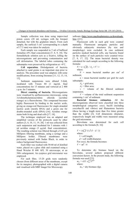
Sample collection was done using improvised<br />
piston corers (20 mL syringes with the forepart<br />
<strong>de</strong>tached, but with the gradation intact). From each<br />
sample, the surficial 5 cm 3 (corresponding to a <strong>de</strong>pth<br />
of 17.5 mm) was taken for analysis.<br />
Each sample was suspen<strong>de</strong>d in 5 ml of buffered<br />
formaline (4% final concentration) [7, 8, 9, 10]. The<br />
formal<strong>de</strong>hy<strong>de</strong> solution acts as a fixative, killing the<br />
microorganisms and preventing contamination and<br />
cell <strong>de</strong>formation. The labeled tubes containing the<br />
subsamples were preserved by refrigeration at +4°C.<br />
Cell separation. Dislodgement of bacteria<br />
attached to sand grains is an important step prior to<br />
analysis. The procedure used was adapted, with some<br />
modifications, from existing literature [11, 12, 13, 14,<br />
15].<br />
Sediment suspensions were diluted 5-fold,<br />
incubated with Tween 80 (1 mg/mL final<br />
concentration) for 15 minutes and vortexed at 2 400<br />
r.p.m. for 5 minutes.<br />
Direct counting of bacteria. Microorganisms<br />
were visualised by epifluorescence microscopy, using<br />
3,6-dimethylaminoacridinic chlori<strong>de</strong> (acridine<br />
orange) as a fluorochrome. This compound becomes<br />
highly fluorescent by binding to the nucleic acids,<br />
giving an orange-red fluorescence for single-stran<strong>de</strong>d<br />
nucleic acids (mostly RNA) and a green one for<br />
double-stran<strong>de</strong>d acids (DNA) [16]. Acridine orange<br />
stains both living and <strong>de</strong>ad cells [17, 18].<br />
The technique employed was an adapted and<br />
simplified version of the protocols used by other<br />
authors [3, 8, 10, 11, 19, 20]. 1 ml was collected from<br />
each suspension and incubated for 5 minutes with 1<br />
ml acridine orange (5 µg/mL final concentration).<br />
The resulting solution was filtered through a 0.45 µm<br />
Millipore filtering membrane, using a syringe and a<br />
Millipore hol<strong>de</strong>r. Filtered membranes were<br />
previously stained with Sudan Black, in or<strong>de</strong>r to<br />
reduce background fluorescence.<br />
Each filter was washed with 50-60 ml of distilled<br />
water, placed on a glass sli<strong>de</strong> and examined using a<br />
Hund Wetzlar H 600 AFL 50 microscope, at an<br />
500× enlargement. An eyepiece grid micrometer was<br />
employed.<br />
For each filter, 15-20 grids were randomly<br />
chosen (from different areas of the membrane, except<br />
for its margins), photographed with a digital camera<br />
and visualised with MBF ImageJ for Microscopy<br />
140<br />
software (http://www.macbiophotonics.ca/downloads.<br />
htm.) [21].<br />
Fluorescent cells in each grid were counted<br />
manually. Fluorescent anorganic particles and<br />
obviously eukaryotic structures (by size and<br />
morphology) were exclu<strong>de</strong>d. In case sediment<br />
particles masked bacterial cells, any bacteria found<br />
on the surface of such particles were counted twice<br />
[7, 8, 17, 22]. The mean bacterial <strong>de</strong>nsity was<br />
calculated for each sample according to the following<br />
formula:<br />
N = n ×Af / Ag × V / v<br />
where:<br />
N = mean bacterial number per cm 3 of<br />
sediment;<br />
n = mean bacterial number per grid for each<br />
subsample;<br />
Ag = grid area;<br />
Af = filter area;<br />
v = volume of the filtered sediment<br />
suspension;<br />
V = volume of the total sediment suspension<br />
containing 1 cm 3 of sediment.<br />
Bacterial biomass estimation. All the<br />
microorganisms observed were classified into three<br />
morphological categories: cocci, bacilli (including<br />
coccobacilli and vibrios) and filamentous bacteria<br />
(those having a length more than five times greater<br />
than the width) [3]. Cell dimensions (diameter,<br />
respectively length and width) were measured using<br />
the grid micrometer.<br />
Biovolume was <strong>de</strong>termined for each cell<br />
according to the formula [8, 16]:<br />
V = (π/ 4) d 2 (l – d / 3)<br />
where:<br />
l = cell length;<br />
d = cell width/diameter.<br />
For cocci, the formula becomes:<br />
V = πd 3 / 6<br />
To <strong>de</strong>termine dry biomass based on the<br />
biovolume, several authors proposed different<br />
conversion factors. In the present study, the following<br />
formula was used [23]:<br />
md = 435 × V 0,86<br />
where:<br />
md = dry biomass (fg);