VOLUM OMAGIAL - Facultatea de Ştiinţe ale Naturii şi Ştiinţe Agricole
VOLUM OMAGIAL - Facultatea de Ştiinţe ale Naturii şi Ştiinţe Agricole
VOLUM OMAGIAL - Facultatea de Ştiinţe ale Naturii şi Ştiinţe Agricole

You also want an ePaper? Increase the reach of your titles
YUMPU automatically turns print PDFs into web optimized ePapers that Google loves.
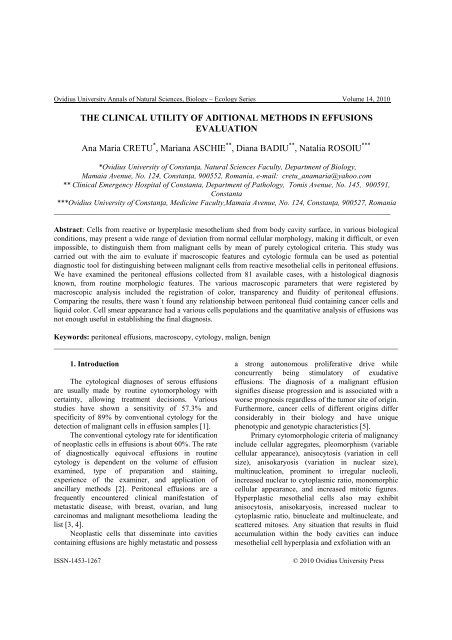
Ovidius University Annals of Natural Sciences, Biology – Ecology Series Volume 14, 2010<br />
THE CLINICAL UTILITY OF ADITIONAL METHODS IN EFFUSIONS<br />
EVALUATION<br />
Ana Maria CRETU * , Mariana ASCHIE ** , Diana BADIU ** , Natalia ROSOIU ***<br />
*Ovidius University of Constanţa, Natural Sciences Faculty, Department of Biology,<br />
Mamaia Avenue, No. 124, Constanţa, 900552, Romania, e-mail: cretu_anamaria@yahoo.com<br />
** Clinical Emergency Hospital of Constanta, Department of Pathology, Tomis Avenue, No. 145, 900591,<br />
Constanta<br />
***Ovidius University of Constanţa, Medicine Faculty,Mamaia Avenue, No. 124, Constanţa, 900527, Romania<br />
________________________________________________________________________________________<br />
Abstract: Cells from reactive or hyperplasic mesothelium shed from body cavity surface, in various biological<br />
conditions, may present a wi<strong>de</strong> range of <strong>de</strong>viation from normal cellular morphology, making it difficult, or even<br />
impossible, to distinguish them from malignant cells by mean of purely cytological criteria. This study was<br />
carried out with the aim to evaluate if macroscopic features and cytologic formula can be used as potential<br />
diagnostic tool for distinguishing between malignant cells from reactive mesothelial cells in peritoneal effusions.<br />
We have examined the peritoneal effusions collected from 81 available cases, with a histological diagnosis<br />
known, from routine morphologic features. The various macroscopic parameters that were registered by<br />
macroscopic analysis inclu<strong>de</strong>d the registration of color, transparency and fluidity of peritoneal effusions.<br />
Comparing the results, there wasn`t found any relationship between peritoneal fluid containing cancer cells and<br />
liquid color. Cell smear appearance had a various cells populations and the quantitative analysis of effusions was<br />
not enough useful in establishing the final diagnosis.<br />
Keywords: peritoneal effusions, macroscopy, cytology, malign, benign<br />
__________________________________________________________________________________________<br />
1. Introduction<br />
The cytological diagnoses of serous effusions<br />
are usually ma<strong>de</strong> by routine cytomorphology with<br />
certainty, allowing treatment <strong>de</strong>cisions. Various<br />
studies have shown a sensitivity of 57.3% and<br />
specificity of 89% by conventional cytology for the<br />
<strong>de</strong>tection of malignant cells in effusion samples [1].<br />
The conventional cytology rate for i<strong>de</strong>ntification<br />
of neoplastic cells in effusions is about 60%. The rate<br />
of diagnostically equivocal effusions in routine<br />
cytology is <strong>de</strong>pen<strong>de</strong>nt on the volume of effusion<br />
examined, type of preparation and staining,<br />
experience of the examiner, and application of<br />
ancillary methods [2]. Peritoneal effusions are a<br />
frequently encountered clinical manifestation of<br />
metastatic disease, with breast, ovarian, and lung<br />
carcinomas and malignant mesothelioma leading the<br />
list [3, 4].<br />
Neoplastic cells that disseminate into cavities<br />
containing effusions are highly metastatic and possess<br />
a strong autonomous proliferative drive while<br />
concurrently being stimulatory of exudative<br />
effusions. The diagnosis of a malignant effusion<br />
signifies disease progression and is associated with a<br />
worse prognosis regardless of the tumor site of origin.<br />
Furthermore, cancer cells of different origins differ<br />
consi<strong>de</strong>rably in their biology and have unique<br />
phenotypic and genotypic characteristics [5].<br />
Primary cytomorphologic criteria of malignancy<br />
inclu<strong>de</strong> cellular aggregates, pleomorphism (variable<br />
cellular appearance), anisocytosis (variation in cell<br />
size), anisokaryosis (variation in nuclear size),<br />
multinucleation, prominent to irregular nucleoli,<br />
increased nuclear to cytoplasmic ratio, monomorphic<br />
cellular appearance, and increased mitotic figures.<br />
Hyperplastic mesothelial cells also may exhibit<br />
anisocytosis, anisokaryosis, increased nuclear to<br />
cytoplasmic ratio, binucleate and multinucleate, and<br />
scattered mitoses. Any situation that results in fluid<br />
accumulation within the body cavities can induce<br />
mesothelial cell hyperplasia and exfoliation with an<br />
ISSN-1453-1267 © 2010 Ovidius University Press