VOLUM OMAGIAL - Facultatea de Ştiinţe ale Naturii şi Ştiinţe Agricole
VOLUM OMAGIAL - Facultatea de Ştiinţe ale Naturii şi Ştiinţe Agricole
VOLUM OMAGIAL - Facultatea de Ştiinţe ale Naturii şi Ştiinţe Agricole

Create successful ePaper yourself
Turn your PDF publications into a flip-book with our unique Google optimized e-Paper software.
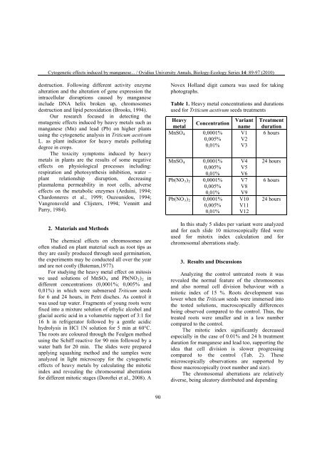
Cytogenetic effects induced by manganese... / Ovidius University Annals, Biology-Ecology Series 14: 89-97 (2010)<br />
<strong>de</strong>struction. Following different activity enzyme<br />
alteration and the alteration of gene expression the<br />
intracellular disruptions caused by manganese<br />
inclu<strong>de</strong> DNA helix broken up, chromosomes<br />
<strong>de</strong>struction and lipid peroxidation (Brooks, 1994).<br />
Our research focused in <strong>de</strong>tecting the<br />
mutagenic effects induced by heavy metals such as<br />
manganese (Mn) and lead (Pb) on higher plants<br />
using the cytogenetic analysis in Triticum aestivum<br />
L. as plant indicator for heavy metals polluting<br />
<strong>de</strong>gree in crops.<br />
The toxicity symptoms induced by heavy<br />
metals in plants are the results of some negative<br />
effects on physiological processes including:<br />
respiration and photosynthesis inhibition, water –<br />
plant relationship disruption, <strong>de</strong>creasing<br />
plasm<strong>ale</strong>ma permeability in root cells, adverse<br />
effects on the metabolic enzymes (Arduini, 1994;<br />
Chardonneres et al., 1999; Ouzounidou, 1994;<br />
Vangronsveld and Clijsters, 1994; Vennitt and<br />
Parry, 1984).<br />
2. Materials and Methods<br />
The chemical effects on chromosomes are<br />
often studied on plant material such as root tips as<br />
they are easily produced through seed germination,<br />
the experiments may be conducted all over the year<br />
and are not costly (Bateman,1977).<br />
For studying the heavy metal effect on mitosis<br />
we used solutions of MnSO4 and Pb(NO3)2 in<br />
different concentrations (0,0001%; 0,005% and<br />
0,01%) in which were submersed Triticum seeds<br />
for 6 and 24 hours, in Petri disches. As control it<br />
was used tap water. Fragments of young roots were<br />
fixed into a mixture solution of ethylic alcohol and<br />
glacial acetic acid in a volumetric rapport of 3:1 for<br />
16 h in refrigerator followed by a gentle acidic<br />
hydrolysis in HCl 1N solution for 5 min at 60°C.<br />
The roots are coloured through the Feulgen method<br />
using the Schiff reactive for 90 min followed by a<br />
water bath for 20 min. The sli<strong>de</strong>s were prepared<br />
applying squashing method and the samples were<br />
analyzed in light microscopy for the cytogenetic<br />
effects of heavy metals by calculating the mitotic<br />
in<strong>de</strong>x and revealing the chromosomal aberrations<br />
for different mitotic stages (Doroftei et al., 2008). A<br />
90<br />
Novex Holland digit camera was used for taking<br />
photographs.<br />
Table 1. Heavy metal concentrations and durations<br />
used for Triticum aestivum seeds treatments<br />
Heavy<br />
metal<br />
MnSO4<br />
MnSO4<br />
Pb(NO3)2<br />
Pb(NO3)2<br />
Concentration Variant<br />
name<br />
0,0001% V1<br />
0,005% V2<br />
0,01% V3<br />
0,0001%<br />
0,005%<br />
0,01%<br />
0,0001%<br />
0,005%<br />
0,01%<br />
0,0001%<br />
0,005%<br />
0,01%<br />
In this study 5 sli<strong>de</strong>s per variant were analyzed<br />
and for each sli<strong>de</strong> 10 microscopically filed were<br />
used for mitotix in<strong>de</strong>x calculation and for<br />
chromosomal aberrations study.<br />
3. Results and Discussions<br />
V4<br />
V5<br />
V6<br />
V7<br />
V8<br />
V9<br />
V10<br />
V11<br />
V12<br />
Treatment<br />
duration<br />
6 hours<br />
24 hours<br />
6 hours<br />
24 hours<br />
Analyzing the control untreated roots it was<br />
reve<strong>ale</strong>d the normal feature of the chromosomes<br />
and also normal cell division behaviour with a<br />
mitotic in<strong>de</strong>x of 15 %. Roots <strong>de</strong>velopment was<br />
lower when the Triticum seeds were immersed into<br />
the tested solutions, macroscopically differences<br />
being observed compared to the control. Thus, the<br />
treated roots were smaller and in a low number<br />
compared to the control.<br />
The mitotic in<strong>de</strong>x significantly <strong>de</strong>creased<br />
especially in the case of 0.01% and 24 h treatment<br />
duration for manganese and lead too, supporting the<br />
i<strong>de</strong>a that cell division is slower progressing<br />
compared to the control (Tab. 2). These<br />
microscopically observations are supported by<br />
those macroscopically (root number and size).<br />
The chromosomal aberrations are relatively<br />
diverse, being <strong>ale</strong>atory distributed and <strong>de</strong>pending