here - Australian College of Veterinary Scientists
here - Australian College of Veterinary Scientists
here - Australian College of Veterinary Scientists

You also want an ePaper? Increase the reach of your titles
YUMPU automatically turns print PDFs into web optimized ePapers that Google loves.
Introduction:<br />
10<br />
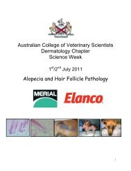
Equine Atopic Dermatitis<br />
ACVSC Proceedings Dermatology Chapter Science Week 2005<br />
Janet Littlewood<br />
Atopic dermatitis is defined as a genetically programmed disease mediated by<br />
reaginic-antibodies (type I hypersensitivity) in which the patient becomes sensitised<br />
to environmental allergens that are innocuous for non-atopic animals. Classically the<br />
diseases is associated with IgE allergen-specific antibodies, but in some species<br />
evidence supports a role for allergen-specific IgG. The importance <strong>of</strong> other<br />
components <strong>of</strong> the immune system are now known to be important in the aetiology<br />
<strong>of</strong> the disease process.<br />
In humans and dogs t<strong>here</strong> is good evidence that the disease is genetically<br />
programmed and t<strong>here</strong> are reports <strong>of</strong> familial incidence and breed predispositions in<br />
the horse that suggest genetic involvement. It is well recognised that insect bite<br />
hypersensitivity (IBH, Culicoides hypersensitivity) has a strong familial tendency, and<br />
t<strong>here</strong> are reports <strong>of</strong> familial occurrence <strong>of</strong> equine atopy suggesting a genetic<br />
predisposition (Rees 2001). Factors such as susceptibility to sensitisation during early<br />
life and the influence <strong>of</strong> the presence <strong>of</strong> parasitic infestation, viral infections,<br />
vaccination and environmental pollutants are <strong>of</strong> unknown significance in the horse.<br />
Genetically-predisposed individuals mount allergen-specific IgE (and possibly IgG)<br />
antibody responses to environmental (and possibly ingested) antigens. Equine IgE<br />
was isolated and characterised by Suter and colleagues in the early 1980's, with even<br />
earlier documentation <strong>of</strong> skin-sensitising antibodies. IgE binds to mast cells in the<br />
skin (and elsew<strong>here</strong>) via the high-affinity IgE receptor. Cross-linking <strong>of</strong> mast cellbound<br />
IgE by specific allergens, causes degranulation and release <strong>of</strong> preformed<br />
mediators, giving rise to the classical erythematous wheal reaction, and synthesis <strong>of</strong><br />
new mediators resulting in the recruitment <strong>of</strong> inflammatory cells to the area.<br />
However, the lesions that develop in atopic humans and dogs at the site <strong>of</strong> patch<br />
tests more closely mimic those found in the naturally occurring clinical disease.<br />
Atopic individuals show increased numbers <strong>of</strong> both dermal and epidermal<br />
Langerhans cells, as well as increased numbers <strong>of</strong> other inflammatory cells.<br />
Epidermal Langerhans cells have been shown to bear allergen-specific IgE molecules<br />
in atopic humans and dogs, further supporting that percutaneous absorption <strong>of</strong><br />
allergens is important in the aetiology <strong>of</strong> the natural disease, probably aided by<br />
epidermal barrier function defects.