here - Australian College of Veterinary Scientists
here - Australian College of Veterinary Scientists
here - Australian College of Veterinary Scientists

Create successful ePaper yourself
Turn your PDF publications into a flip-book with our unique Google optimized e-Paper software.
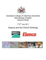
Introduction<br />
Diagnostic Approach to Urticaria<br />
Reg Pascoe<br />
Horses show the greatest incidence <strong>of</strong> urticaria <strong>of</strong> all the species <strong>of</strong> domestic animals.<br />
Urticaria is a specific skin lesion rather than a specific disease entity. It has many<br />
different aetiologies and pathogenesis (Logas & Barbet 1999). Generally, it is discussed<br />
as a single entity even though its clinical manifestations vary from a minor transitory<br />
nature to major systemic life endangering problems.<br />
Aetiology / Pathophysiology<br />
The pathogenesis <strong>of</strong> equine urticaria is not well understood. Urticaria has been<br />
associated with immunological and nonimmunological mechanisms leading to the<br />
release <strong>of</strong> various mediators by mast cells. In a study by Rufnacht et al (2004) skin<br />
biopsies <strong>of</strong> 32 horses with a history <strong>of</strong> urticaria were stained with toluidine blue, a<br />
double-labelling method for chymase and tryptase, plus immunohistochemistry for<br />
immunoglobulin IgE. These horses were compared with horses with pemphigus<br />
foliaceus, insect bite hypersensitivity and control horses with healthy skin. Neither<br />
formalin fixation time nor biopsy site influenced the staining methods.No chymasepositive<br />
cells were found. In all groups <strong>of</strong> horses, cells staining with toluidine blue and<br />
for tryptase and IgE were found in the epidermis and hair follicle papilla and<br />
significantly more positively staining cells were observed in the subepidermal dermis<br />
compared with the deep dermis. Horses with urticaria had significantly more IgEbearing<br />
cells in the subepidermal dermis than control horses. However, horses with<br />
urticaria had significantly fewer toluidine-blue-stained mast cells in both subepidermal<br />
and deep dermis compared with the insect bite hypersensitivity and pemphigus<br />
foliaceus groups.<br />
This study supports IgE-mediated reactions play a role in the pathogenesis <strong>of</strong> urticaria.<br />
The use <strong>of</strong> intradermal testing (IDT) <strong>of</strong> urticaria and atopy also can be equivocal as<br />
Evans et al (1992) had found that positive results were also obtained from horses not<br />
showing signs <strong>of</strong> atopy. Further studies (Lorch et al 2001) have assisted in the<br />
elucidation <strong>of</strong> more <strong>of</strong> the quandary associated with IDT<br />
Many causes <strong>of</strong> urticaria have been suggested. (Scott & Miller 2003)<br />
1. Degranulation <strong>of</strong> mast cells and basophils is presumed to be the basic<br />
pathogenesis. Liberation <strong>of</strong> chemical mediators, which cause increased vascular<br />
permeability leads to wheal formation.<br />
ACVSC Proceedings Dermatology Chapter Science Week 2005 41