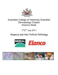
here - Australian College of Veterinary Scientists
here - Australian College of Veterinary Scientists
here - Australian College of Veterinary Scientists

Create successful ePaper yourself
Turn your PDF publications into a flip-book with our unique Google optimized e-Paper software.
fibroblastic variant, particularly on the elbow or jaw. Differential diagnoses for such<br />
lesions include various forms <strong>of</strong> chronic infectious lymphangitis/lymphadenitis (e.g.<br />
glanders, epizootic lymphangitis and cutaneous histoplasmosis) and metastasising<br />
carcinomas.<br />
Why biopsy?<br />
Histopathology usually permits confirmation <strong>of</strong> a clinical diagnosis <strong>of</strong> equine sarcoid<br />
and elimination <strong>of</strong> the considerable list <strong>of</strong> differential diagnoses. However, nonexcisional<br />
biopsy may provoke transition <strong>of</strong> a quiescent sarcoid into a rapidly<br />
growing aggressive one. For this reason, many equine practitioners are loath to<br />
interfere, particularly with large or poorly demarcated lesions or when a suitable<br />
method <strong>of</strong> treatment is unlikely to be available once the diagnosis is confirmed.<br />
Diagnostic histological features <strong>of</strong> equine sarcoids<br />
Histologically, most sarcoids are characterised by dermal fibroblastic proliferation<br />
and intimately associated, pseudoepitheliomatous hyperplasia <strong>of</strong> the epidermis.<br />
T<strong>here</strong> is usually some degree <strong>of</strong> hyperkeratosis and t<strong>here</strong> may be mild parakeratosis.<br />
Long, narrow, anastomosing rete pegs <strong>of</strong> hyperplastic and at least focally acanthotic<br />
epidermis extend into the proliferating spindle cell component in the dermis.<br />
The infiltrative and poorly demarcated dermal component is usually <strong>of</strong> moderately<br />
high cell density, with the superficial portion tending to be more cellular than the<br />
deep part. The dermal fibroblasts are slender to plump fusiform cells with poorly<br />
defined cytoplasmic boundaries. The nuclei may be slender and elongate or enlarged<br />
and <strong>of</strong> irregular shape. Mitotic figures are usually infrequent (0-1 per high power<br />
field) but may be more numerous (up to 2-3 per high power field) in superficial than<br />
in deep areas <strong>of</strong> sarcoids and adjacent to areas <strong>of</strong> ulceration. Mitoses and<br />
fibroblastic nuclear pleomorphism, anisokaryosis and nucleolar enlargement may be<br />
observed in rapidly growing or recurrent sarcoids; cellular anaplasia is, however, rare.<br />
The fibroblasts are typically arrayed in tight whorls, interweaving bundles, haphazard<br />
tangles and occasionally herringbone and basket weave patterns, with a small to<br />
moderate amount <strong>of</strong> intercellular collagen. In recently developed sarcoids, the<br />
volume <strong>of</strong> intercellular collagen may be minimal. In chronic lesions, the collagen<br />
content increases and the dense fibres may appear hyalinised. Plump dermal<br />
fibroblasts usually directly abut the epidermis and, in a high percentage <strong>of</strong> sarcoids,<br />
are characteristically oriented perpendicular to the epidermal basement membrane in<br />
a picket-fence arrangement. Hair follicles enveloped by fibroblasts may undergo<br />
cystic dilation.<br />
Histological variants<br />
Prominent hyperplasia <strong>of</strong> the epidermis with formation <strong>of</strong> long rete pegs is not<br />
invariably present in sarcoids. It is usually observed in verrucous and mixed forms<br />
and at the margins <strong>of</strong> ulcerated areas in fibroblastic sarcoids. In occult sarcoids, the<br />
80<br />
ACVSC Proceedings Dermatology Chapter Science Week 2005