here - Australian College of Veterinary Scientists
here - Australian College of Veterinary Scientists
here - Australian College of Veterinary Scientists

Create successful ePaper yourself
Turn your PDF publications into a flip-book with our unique Google optimized e-Paper software.
Introduction<br />
100<br />
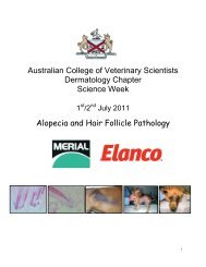
How I treat: equine phycomycosis<br />
ACVSC Proceedings Dermatology Chapter Science Week 2005<br />
Reg Pascoe<br />
Chronic, subcutaneous, fungal, ulcerative, granulomatous, subtropical and tropical<br />
skin disease caused by Pythium insidiosum. Affects horses <strong>of</strong> all breeds, ages and sexes.<br />
Most cases occur in summer and autumn Other names include Bursatee, Florida<br />
horse leech, Swamp cancer.(Bridges & Emmons 1961, Blackford 1984 Scott & Miller<br />
2003)<br />
Aetiology / Pathophysiology<br />
Caused by Pythium spp., free-living aquatic organisms which are not true fungi. Horses<br />
become infected by standing for long periods in stagnant water with rotting organic<br />
material and high ambient temperatures. 30º C to 40º C favour infection. Damaged<br />
skin assists the entry <strong>of</strong> the organisms into the horse’s skin<br />
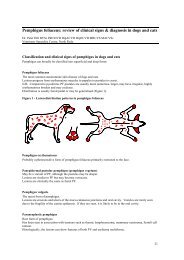
Clinical Presentation<br />
T<strong>here</strong> is no age, sex or breed predeliction. Pruritus occurs early in the infection with<br />
biting and kicking at affected area with subsequent ulceration <strong>of</strong> skin or wound<br />
Most lesions occur on legs and lower abdomen and chest. Lesions are usually single<br />
and unilateral but occasionally are multiple. Body lesions are <strong>of</strong>ten roughly circular<br />
with a sticky, serosanguineous, stringy discharge which either mats hairs or hangs<br />
from body wall in thick mucopurulent strands. Numerous irregular, gritty,coral like<br />
bodies (kunkers) occur within the necrotic sinuses found in the lesion. These are<br />
composed <strong>of</strong> fungal hyphae and tissue debris. Involvement <strong>of</strong> joint and tendons with<br />
sinus formation is a serious complication. Bone involvement is seldom seen in<br />
lesions less than four weeks old. Chronically affected horses may show invasion <strong>of</strong><br />
underlying bone commonest areas are the cannon bones sesamoids and phalanges<br />
Lymphadenopathy occurs in chronic cases<br />
Differential diagnosis<br />
Sarcoid, habronema infestation in wounds, neoplasms (especially sarcoids and<br />
squamous cell carcinomas), Mycetoma, Botryomycosis, excess granulation tissue,<br />
foreign body granulomas and other Zygomycetes.