here - Australian College of Veterinary Scientists
here - Australian College of Veterinary Scientists
here - Australian College of Veterinary Scientists

You also want an ePaper? Increase the reach of your titles
YUMPU automatically turns print PDFs into web optimized ePapers that Google loves.
y a variety <strong>of</strong> fungal metabolites. T equinum produces urease, gelatinase, protease,<br />
hemolysins, keratinases and lipases. Trichophyton spp<br />
have been shown to produce proteolytic enzymes capable <strong>of</strong> inducing acantholysis.<br />
As well as pro-inflammatory metabolites T. mentagrophytes produces mannans that<br />
may inhibit T cell responses and reduce epidermal turnover (exfoliation being an<br />
innate defence mechanism <strong>of</strong> the skin to surface microbes). Concurrent bacterial<br />
infection is sometimes seen in dermatophytosis and dermatophytes may liberate<br />
penicillin-like substances that result in the formation <strong>of</strong> penicillin resistant bacteria<br />
from affected skin.<br />
T<strong>here</strong> seems to be a lack <strong>of</strong> specific immunological studies in the equine however<br />
based on other species it would appear that spontaneous resolution is associated<br />
with the development <strong>of</strong> cell mediated immunity (IFNγ and IL2). T<strong>here</strong> does not<br />
appear to be any correlation with antibody titres. Recovered horses from T equinum<br />
and M. gypseum infections are reported to be resistant to re-infection with<br />
dermatophytes <strong>of</strong> the same species.<br />
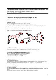
Clinical features:<br />
Dermatophytosis is a follicular infection so the common clinical symptom is <strong>of</strong><br />
folliculitis. This may include hive like follicular tufts, annular, peripherally expanding<br />
areas <strong>of</strong> alopecia with variable scaling or crusting, classical ring lesion with peripheral<br />
papules, coalescing areas forming serpiginous patterns <strong>of</strong> alopecia, exudative lesions<br />
on the caudal fetlocks “grease heal” and rarely subcutaneous and dermal nodules<br />
(dermatophytic pseudomycetoma). Pruritus is minimal or absent unless complicated<br />
by ectoparasites and pain is uncommon unless complicated by Staphylococcal<br />
folliculitis.<br />
Lesions in dermatophytosis are most commonly seen on the face, neck, dorsolateral<br />
thorax and the girth area. It has been reported that dermatophytosis affecting the<br />
girth is most likely to be T equinum (fomite transmission) and that M. gypseum is most<br />
commonly isolated from face, neck, legs and rump (geophylic fungus spread via<br />
arthropods). T verrucosum is reported to have a thicker, grayish crust. Due to clinical<br />
overlap, lesion type and location should not be relied on to identify species <strong>of</strong><br />
dermatophyte involved.<br />
Zoonotic factors:<br />
T equinum, T. verrucosum and M. canis all have zoonotic potential<br />
Differential diagnoses:<br />
Other folliculitides<br />
- Staphylococcal folliculitis<br />
- Dermatophilosis<br />
- Pemphigus foliaceous<br />
- Eosinophilic folliculitis<br />
ACVSC Proceedings Dermatology Chapter Science Week 2005 107