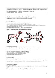
here - Australian College of Veterinary Scientists
here - Australian College of Veterinary Scientists
here - Australian College of Veterinary Scientists

Create successful ePaper yourself
Turn your PDF publications into a flip-book with our unique Google optimized e-Paper software.
the pathology labs was for granulomatous skin. Recently, a group <strong>of</strong> 7 horses were<br />
investigated which had primary pulmonary disease and no reported concurrent<br />
dermatologic symptoms (Pusterla and others 2003). All <strong>of</strong> these horses showed<br />
granulomatous lesions in the lungs, which formed the inclusion criterion for the<br />
report. Cutaneous signs may not have been present, not mentioned specifically or<br />
overlooked due to the gravity <strong>of</strong> the systemic disease. T<strong>here</strong> may be significant<br />
similarity between human and equine sarcoidosis and the disease may present with a<br />
spectrum <strong>of</strong> clinical signs. Dermatologic lesions may be on one end <strong>of</strong> the spectrum<br />
and pulmonary lesions on the other.<br />
Sarcoidosis is a rare disease in equine medicine and the pathogenesis is unclear. In<br />
human medicine, histopathologic findings <strong>of</strong> sarcoidosis resemble those seen in<br />
tuberculosis. Furthermore, tuberculosis shows an increased incidence in patients<br />
with sarcoidosis. As long as 35 years ago, a role for atypical mycobacteria was<br />
presented (Mankiewicz 1964). By using molecular biologic methods, DNA from M.<br />
tuberculosis and an unidentified nontuberculous mycobacterium was identified in<br />
tissue sections and bronchial washings <strong>of</strong> some patients with sarcoidosis (Fidler and<br />
others 1993). Multiple species <strong>of</strong> mycobacteria were detected by polymerase chain<br />
reaction (PCR) predominantly from samples <strong>of</strong> the lymph nodes and pulmonary<br />
tissue (Li and others 1999). These findings support a potential role <strong>of</strong> mycobacteria<br />
in the pathogenesis <strong>of</strong> sarcoidosis in some human patients. However it must not be<br />
thought to be the exclusive infectious agent as recent studies in Japan and Europe<br />
suggest that Propionibacterium spp. may also be involved in the aetiology <strong>of</strong> sarcoidosis<br />
(Eishi and others 2002). Infectious agents could not be detected in horses by culture,<br />
electron microscopy, animal inoculation, direct immun<strong>of</strong>luorescence or<br />
immunoperoxidase testing (Heath and others 1990, Scott 1991, Stannard 1987, von<br />
Tscharner and others 2000). In three previously reported cases positive titers to<br />
Borrelia burgdorferi were shown (Rose and others 1996). In a recent report, paraffinembedded<br />
skin specimens from 8 horses with sarcoidosis were evaluated with special<br />
stains and PCR assays for Mycobacteria spp., Borrelia burgdorferi, Coccidioides immitis,<br />
Cryptococcus ne<strong>of</strong>ormans and Corynebacterium pseudotuberculosis to no avail (Spiegel and<br />
others 2005). T<strong>here</strong> are anecdotal reports about improvement <strong>of</strong> the condition in<br />
horses which were moved from one state to another (personal communication Tony<br />
Stannard). It was hypothesized that the beneficial effect <strong>of</strong> relocation was due to<br />
differences in environmental concentrations <strong>of</strong> mould spores which may play a role<br />
in the pathogenesis <strong>of</strong> the disease. None <strong>of</strong> the horses included in this study had a<br />
correspondent history.<br />
In humans, massive immunosuppressive doses <strong>of</strong> glucocorticoids are the treatment<br />
<strong>of</strong> choice for sarcoidosis with significant systemic involvement (Braverman 2003).<br />
Cutaneous lesions generally respond to therapy for systemic involvement, but <strong>of</strong>ten<br />
recur when the dosage is lowered or discontinued. Patients without systemic<br />
involvement are typically not treated with glucocorticoids. T<strong>here</strong> are anecdotal<br />
reports <strong>of</strong> success with chloroquine (Zic and others 1991), hydroxychloroquine<br />
(Jones and others 1990), methotrexate (Lower and others 1995), minocycline<br />
(Bachelez and others 2001), thalidomide (Calderon and others 1997, English and<br />
62<br />
ACVSC Proceedings Dermatology Chapter Science Week 2005