here - Australian College of Veterinary Scientists
here - Australian College of Veterinary Scientists
here - Australian College of Veterinary Scientists

You also want an ePaper? Increase the reach of your titles
YUMPU automatically turns print PDFs into web optimized ePapers that Google loves.
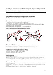
any anatomic location but commonly arise on the medial thigh, prepuce, neck and<br />
face.<br />
Verrucous (warty) sarcoids are usually less circumscribed than occult sarcoids and<br />
protrude as palpable masses due to an increased dermal neoplastic component.<br />
They may be sessile or pedunculated. The degree <strong>of</strong> overlying hyperkeratosis and<br />
scaling is variable but t<strong>here</strong> is <strong>of</strong>ten a partially or totally alopecic, dry, cauliflower-like<br />
surface. This variant is also relatively slow-growing but a more aggressive<br />
fibroblastic type may emerge following trauma. Verrucous sarcoids need to be<br />
distinguished from squamous papillomas induced by equine papillomavirus and from<br />
chronic frictional irritation. Verrucous sarcoids are most commonly located in the<br />
axillary and inguinal areas.<br />
Fibroblastic sarcoids involve both the dermis and subcutis, with the extent <strong>of</strong> deep<br />
dermal and subcutaneous infiltration <strong>of</strong>ten being underestimated on clinical<br />
examination. This variant typically manifests as a protruberant, firm to fleshy,<br />
ulcerated mass which closely resembles exuberant granulation tissue. It is wellvascularised<br />
and prone to bleed when minimally traumatised. Most sarcoids arising<br />
on the brisket or limbs are <strong>of</strong> fibroblastic appearance. Fibroblastic sarcoids may<br />
enlarge rapidly over weeks to months and then stabilise and persist for years.<br />
Differential diagnoses for such lesions include, in addition to proud flesh, squamous<br />
cell carcinoma, botryomycosis, habronemiasis, phycomycosis, fibrosarcoma and<br />
peripheral nerve sheath tumour.<br />
Nodular sarcoids usually involve only the subcutis as one or multiple, spherical,<br />
mobile nodules usually covered by intact skin <strong>of</strong> normal appearance. Intradermal<br />
nodular sarcoids may occasionally be seen and the overlying skin may appear thin<br />
and shiny. Sarcoids involving the palpebral margins are typically nodular and this<br />
variant is also commonly found on the medial thigh, prepuce and inguinal area.<br />
Nodular sarcoids, being well demarcated, are amenable to surgical excision but<br />
incomplete excision (or traumatic ulceration <strong>of</strong> the overlying skin) may encourage<br />
emergence <strong>of</strong> a more aggressive fibroblastic variant. Nodular sarcoids may grossly<br />
resemble nodular necrobiosis (equine eosinophilic collagenolytic granuloma) and<br />
other mesenchymal tumours such as melanoma, fibroma or peripheral nerve sheath<br />
tumour.<br />
Mixed sarcoids present with a combination <strong>of</strong> the gross characteristics <strong>of</strong> two or<br />
more <strong>of</strong> the verrucous, fibroblastic and nodular forms and may represent a<br />
transitional phase as a more rapidly growing and locally aggressive neoplasm emerges<br />
from a previously stable focus. Mixed sarcoids are most commonly found in the<br />
axillary and inguinal areas and on the face.<br />
Malevolent sarcoids are rare, highly invasive variants which infiltrate along<br />
lymphatics to produce nodular cording and multiple ulcerated fibroblastic nodules.<br />
The neoplastic tissue may extend along lymphatics to involve regional lymph nodes.<br />
Most malevolent sarcoids have been observed following intereference with a<br />
ACVSC Proceedings Dermatology Chapter Science Week 2005 79