here - Australian College of Veterinary Scientists
here - Australian College of Veterinary Scientists
here - Australian College of Veterinary Scientists

Create successful ePaper yourself
Turn your PDF publications into a flip-book with our unique Google optimized e-Paper software.
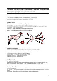
Clinical Disease<br />
The clinical result <strong>of</strong> disruption to blood supply from cutaneous vasculitis is lesions<br />
that range in severity from wheals, papules, purpura, erythema and oedema if acute;<br />
to alopecia and scarring if mild and chronic; to areas <strong>of</strong> well-demarcated necrosis,<br />
ulceration and crusting if severe. Lesions occur most commonly on the distal limbs,<br />
pinnae, lips and periocular areas. Diascopy confirms purpura as erythemic lesions<br />
that will not blanch. Signs <strong>of</strong> systemic illness, including pyrexia, lethargy, decreased<br />
appetite and weight loss may accompany cutaneous lesions. Forms <strong>of</strong> cutaneous<br />
vasculitis recognised in horses include:<br />
1. Purpura Haemorrhagica - is reported as the most common form <strong>of</strong><br />
cutaneous vasculitis in the horse. It is an acute disease characterised by extensive<br />
oedematous swelling <strong>of</strong> the distal limbs, ventral abdomen, and sometimes face and<br />
muzzle, and typically concurrent signs <strong>of</strong> systemic illness. Incidence is low and<br />
sporadic, with most affected horses greater than 2 years <strong>of</strong> age, and no apparent<br />
breed or sex predispositions. This syndrome may occur within 2-4 weeks <strong>of</strong> a<br />
respiratory infection (Streptococcus equi or zooepidemicus), or other potential triggering<br />
factors or disease. Urticaria may occur initially, and progress to oedema that is<br />
usually well-demarcated, painful, symmetrical, and involving all four limbs. Severely<br />
affected areas may have surface serum exudation and progress to necrosis and<br />
ulceration. Petechiae and ecchymoses may be present on mucous membranes. Most<br />
affected horses have obvious signs <strong>of</strong> depression and are reluctant to move,<br />
anorexic, tachycardic, tachypnoeic, and <strong>of</strong>ten pyrexic. T<strong>here</strong> is a wide variation in<br />
severity and clinical course.<br />
2. Subacute Pastern Vasculitis – is proposed to be a relatively common<br />
although poorly characterised entity in the horse. Lesions primarily occur on nonpigmented<br />
lower limbs, and may be more prevalent in animals on pasture. Sunlight<br />
exposure and photactivation are proposed aetiological factors in some cases,<br />
however appear uninvolved in others. Early lesions include erythema and<br />
oedematous swelling, which progress to serum exudation, necrosis, crusting and<br />
ulceration. Pain on palpation is <strong>of</strong>ten present, and pruritus absent. Chronic lesions<br />
may be alopecic, scaling, lichenified, and proliferative.<br />
Diagnosis<br />
Definitive diagnosis <strong>of</strong> cutaneous vasculitis requires consistent history, clinical<br />
lesions, and histopathological changes. In acute disease varying degrees <strong>of</strong> specific<br />
vessel wall changes may be identified, including disproportionately more leukocytes<br />
within vessel walls than surrounding perivascular areas, leukocytoclasis within or<br />
adjacent to walls, fibrinoid necrosis, and vessel thrombosis. Less specific supportive<br />
changes include intense oedema, red cell extravasation, and endothelial cell swelling.<br />
T<strong>here</strong> may be evidence <strong>of</strong> cutaneous infarction if severe, and follicular atrophy<br />
(“faded follicles”) with chronic disease. Clear histological support for a diagnosis <strong>of</strong><br />
ACVSC Proceedings Dermatology Chapter Science Week 2005 113