here - Australian College of Veterinary Scientists
here - Australian College of Veterinary Scientists
here - Australian College of Veterinary Scientists

Create successful ePaper yourself
Turn your PDF publications into a flip-book with our unique Google optimized e-Paper software.
frequently generalizes within 1-3 months (Von Tscharner and others 2000, Scott and<br />
Miller 2003). The coronary band may show changes and may be the only affected<br />
site (Fadok 1995, Von Tscharner and others 2000). Other targets might be the<br />
prepuce or the mammary glands (Johnson 1997, Scott and Miller 2003). Pitting<br />
oedema <strong>of</strong> the lower extremities and the ventral abdomen is seen in over 50% <strong>of</strong> the<br />
cases (Manning 1983, Scott 1989, White 1992) and might be the only initial sign <strong>of</strong><br />
disease (Scott and Manning 1983, Schulte and others 1989, Von Tscharner and<br />
others 2000, Scott and Miller 2003). Urticaria can occur weeks before classical<br />
lesions such as pustules and/or crusts are seen (Von Tscharner and others 2000).<br />
Some authors state that horses may occasionally be pruritic, but more <strong>of</strong>ten are<br />
painful (Von Tscharner and others 2000, Scott and Miller 2003), others mention<br />
pruritus in 50% <strong>of</strong> the horses (Zabel and others 2005).<br />
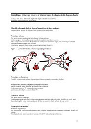
The primary lesions in PF are vesicles, bullae and pustules. These lesions are very<br />
fragile and short-lived. Most <strong>of</strong>ten patients present with secondary lesions such as<br />
erosions, epidermal collarettes, crusts, alopecia, and scaling (Von Tscharner and<br />
others 2000, Scott and Miller 2003). Systemic signs like fever, depression, anorexia,<br />
lethargy and weight loss (Scott 1989, White 1992, Manning 1983), and nonregenerative<br />
anaemia, neutrophilia, hypoalbuminia, elevated alkaline phosphatase,<br />
elevated fibrinogen and hyperglobunemia may occur (George 1984, Edmond 1986,<br />
Day and others 1986, Scott and others 1987).<br />
Tests useful in the diagnosis <strong>of</strong> equine pemphigus foliaceus include direct smears,<br />
skin biopsies and immun<strong>of</strong>luorescence or immunohistochemical testing (Scott and<br />
Miller 2003). Cytology from crusted areas or intact pustules may show large<br />
numbers <strong>of</strong> neutrophils and acantholytic cells and is reported to be a useful test in<br />
the diagnosis <strong>of</strong> equine pemphigus foliaceus. Multiple biopsies from intact pustules<br />
or crusts are needed to confirm the diagnosis (Von Tscharner and others 2000).<br />
Primary dermatohistopathologic findings include subcorneal and/or intraepidermal<br />
pustules, associated with marked acantholysis (Scott 1989, Yaeger and Scott 1993,<br />
Von Tscharner and others 2000). In the absence <strong>of</strong> pustules, large numbers <strong>of</strong><br />
acantholytic cells and neutrophils found in the crusts are <strong>of</strong>ten the only diagnostic<br />
finding. As acantholysis may also be seen in dermatophytosis, special stains and<br />
fungal cultures are recommended (Scott and Miller 2003). Histopathology is<br />
reported to be far more reliable than immunopathologic testing in the diagnosis <strong>of</strong><br />
equine pemphigus foliaceus. (Scott and others 1984, Day and Penhale 1986, Griffith<br />
1987, Scott and Miller 2003).<br />
The prognosis for equine pemphigus foliaceus patients depends on age <strong>of</strong> onset.<br />
Younger horses (< 1 year) seem to have a better prognosis, the disease tends to be<br />
less severe, responds better to treatment and may spontaneously regress or not<br />
require further medication once in remission (Laing and others 1992, Von Tscharner<br />
and others 2000, Scott and Miller 2003). However, mature horses (> 5 years) have a<br />
less favourable prognosis and most require aggressive treatment (Von Tscharner and<br />
others 2000). Spontaneous remission is rare, but has been reported (White 1992,<br />
Amory and others 1997, Von Tscharner and others 2000, Scott and Miller 2003).<br />
96<br />
ACVSC Proceedings Dermatology Chapter Science Week 2005