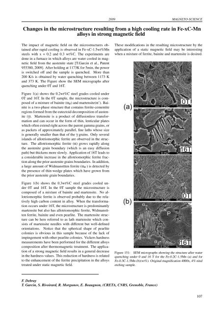
2009 MAGNETO-SCIENCEChanges in the microstructure resulting from a high cooling rate in Fe-xC-Mnalloys in strong magnetic fieldThe impact of magnetic field on the microstructures obtainedafter rapid cooling is observed in Fe-xC-1.5wt%Mnsteels with x = 0.2 and 0.3 wt%C. The experim<strong>en</strong>ts aredone in a furnace in which alloys are water cooled in magneticfield from the aust<strong>en</strong>ite state [T.Garcin et al., Pat<strong>en</strong>t955380, 2009]. After holding at 1173K for 5min, the poweris switched off and the sample is qu<strong>en</strong>ched. More than200 K/s is obtained by water qu<strong>en</strong>ching betwe<strong>en</strong> 1173 Kand 373 K. The Figure show the SEM micrographs afterqu<strong>en</strong>ching under 0T and 16T.These modifications in the resulting microstructure by theapplication of a static magnetic field may be interestingwh<strong>en</strong> a mixture of ferrite, bainite and mart<strong>en</strong>site is <strong>des</strong>ired.Figure 1(a) shows the 0.2wt%C steel gra<strong>des</strong> cooled under0T and 16T. In the 0T sample, the microstructure is composedof a mixture of bainite (α B ) and mart<strong>en</strong>site(α’). Bainiteis a two-phase structure that contains ferrite-cem<strong>en</strong>titeregions formed from the eutectoid decomposition of aust<strong>en</strong>ite(γ). Mart<strong>en</strong>site is a product of diffusionless transformationand can occur in the form of thin, l<strong>en</strong>ticular plateswhich oft<strong>en</strong> ext<strong>en</strong>d right across the par<strong>en</strong>t gamma grains, oras packets of approximately parallel, fine laths whose sizeis g<strong>en</strong>erally smaller than that of the γ grains. Only severalislands of allotriomorphic ferrite are observed in the structure.The allotriomorphic ferrite (α) grows rapidly alongthe aust<strong>en</strong>ite grain boundary (which is an easy diffusionpath) but thick<strong>en</strong>s more slowly. Application of 16T leads toa considerable increase in the allotriomorphic ferrite fractionalong the prior aust<strong>en</strong>ite grains boundaries. In addition,a large amount of Widmansttt<strong>en</strong> ferrite (α W ) is detected bythe pres<strong>en</strong>ce of thin-wedge plates which have grown fromthe prior aust<strong>en</strong>ite grain boundaries.Figure 1(b) shows the 0.3wt%C steel gra<strong>des</strong> cooled under0T and 16T. In the 0T sample the microstructure iscomposed of a mixture of bainite and mart<strong>en</strong>site. No allotriomorphicferrite is observed probably due to the relativelyhigh carbon cont<strong>en</strong>t in alloy. Wh<strong>en</strong> the transformationoccurs under 16T, the microstructure is predominantlymart<strong>en</strong>site but also has allotriomorphic ferrite, Widmansttt<strong>en</strong>ferrite, bainite and ev<strong>en</strong> pearlite. The mart<strong>en</strong>site structurecan be here referred to as lath mart<strong>en</strong>site which consistsof mart<strong>en</strong>site needles with differ<strong>en</strong>t but well-definedori<strong>en</strong>tations. Notice that the spherical shape of pearlitecolonies is obvious in this sample because of the lack ofimpingem<strong>en</strong>t with other pearlite colonies. Vickers hardnessmeasurem<strong>en</strong>ts have be<strong>en</strong> performed for the differ<strong>en</strong>t alloyscomposition after thermomagnetic treatm<strong>en</strong>t. The applicationof a strong magnetic field results in a g<strong>en</strong>eral decreasein the hardness values. This reduction of hardness is relatedto the <strong>en</strong>hancem<strong>en</strong>t of the ferrite precipitation in the alloystreated under static magnetic field.Figure 151: SEM micrographs showing the structure after waterqu<strong>en</strong>ching under 0 and 16 T for the Fe-0.2C-1.5Mn (a) and forFe-0.3C-1.5Mn (b)(wt%). Original magnification 4000x, 4% nitaletching sample.F. DebrayT. Garcin, S. Rivoirard, R. Morgunov, E. Beaugnon, (CRETA, CNRS, Gr<strong>en</strong>oble, France)107
MAGNETO-SCIENCE 2009Study of the influ<strong>en</strong>ce of magnetic forces on the mass transferof paramagnetic particles in electrochemistryThe context of our work is the characterization of the magneticforces which influ<strong>en</strong>ce the processes of mass transferin electrochemistry. It appears experim<strong>en</strong>tally that in thepres<strong>en</strong>ce of a magnetic field in solutions, where a gradi<strong>en</strong>tof conc<strong>en</strong>tration of paramagnetic ions is pres<strong>en</strong>t, ev<strong>en</strong> if theaction of Lor<strong>en</strong>tz forces is not effective, a driving force actingon the solution will arise. This force, referred to as theconc<strong>en</strong>tration gradi<strong>en</strong>t force, is oft<strong>en</strong> writt<strong>en</strong>,−→ B 2−→ ∇CF m = χ m , (18)2µ 0where χ m (m 3 /mol) is the molar magnetic susceptibility,B(T) the magnetic flux d<strong>en</strong>sity, C(mol/m 3 ) the bath conc<strong>en</strong>trationand µ 0 (Hm −1 ) the permeability of vacuum. Theexperim<strong>en</strong>t was performed using a 10 MW resistive magnetwhich g<strong>en</strong>erates a homog<strong>en</strong>eous a magnetic field of 6 T ina 286 mm diameter. The experim<strong>en</strong>tal device is a rectangularchannel with platinum electro<strong>des</strong> on the upper and lowerwalls, betwe<strong>en</strong> which a voltage drop is applied (figure 152).The channel is closed with insulating walls to eliminate theedge effects. The cell is immersed in an electrolytic bathprepared with an equimolar solution of 0.05 mol/m 3 Ferriferro-cyanide with 0.5 mol/m 3 and K 2 SO 4 as the supportingelectrolyte. The temperature condition was about 17 ◦ Cand an Ag/AgCl refer<strong>en</strong>ce electrode was used to control theelectro<strong>des</strong> pot<strong>en</strong>tial.of the boundary layer. As a result, an additional mechanismof transport of the solution is g<strong>en</strong>erated. Correspondingly,the limiting curr<strong>en</strong>ts of reactions proceeding in the electrochemicalsystem will become a function of the appliedmagnetic flux d<strong>en</strong>sity. The influ<strong>en</strong>ce of magnetic field onthe evolution of the mean limiting curr<strong>en</strong>t d<strong>en</strong>sity is plottedusing logarithmic coordinates in figure 153. We can showsthat for the two mo<strong>des</strong>, anodic and cathodic, the limitingcurr<strong>en</strong>t d<strong>en</strong>sity follows a law in B 2/3 . Note that this lawstarts at 0.5 T for the anodic mode and at 2 T for the cathodicmode due to the paramagnetic forces which drivesa more important flow than the gravity. This ph<strong>en</strong>om<strong>en</strong>ologicalbehaviour was observed by many authors [Waskaas,Acta Chimica 50, 516 (1996)], Rabahand et al., Journal ofElectroanalytical Chemistry 571, 85 (2004)].Figure 152:Experim<strong>en</strong>tal configuration.The results were obtained by the polarographic method.Wh<strong>en</strong> the two electro<strong>des</strong> located on the faces of the channelare submitted to a voltage drop, controlled by a pot<strong>en</strong>tiostat,an electric curr<strong>en</strong>t is imposed and in the pres<strong>en</strong>ce ofan homog<strong>en</strong>eous magnetic field, the gradi<strong>en</strong>t of paramagneticions due to electrode reaction will cause a redistributionof velocities in the bath which th<strong>en</strong> acts on the depthFigure 153: Evolution of the limiting curr<strong>en</strong>t d<strong>en</strong>sities with thework electrode (WE) in the (a) cathodic mode and (b) anodicmode.F. DebrayD. Baaziz, A. Alemany, (EPM-SIMAP Laboratory, Gr<strong>en</strong>oble, France), D. Kalache (Fluids Mechanics Laboratory,University of USTHB, Algiers)108
- Page 1 and 2:
LABORATOIRE NATIONAL DES CHAMPS MAG
- Page 4 and 5:
TABLE OF CONTENTSPreface 1Carbon Al
- Page 6 and 7:
Coexistence of closed orbit and qua
- Page 8:
2009PrefaceDear Reader,You have bef
- Page 12 and 13:
2009 CARBON ALLOTROPESInvestigation
- Page 14 and 15:
2009 CARBON ALLOTROPESPropagative L
- Page 16 and 17:
2009 CARBON ALLOTROPESEdge fingerpr
- Page 18 and 19:
2009 CARBON ALLOTROPESObservation o
- Page 20 and 21:
2009 CARBON ALLOTROPESImproving gra
- Page 22 and 23:
2009 CARBON ALLOTROPESHow perfect c
- Page 24 and 25:
2009 CARBON ALLOTROPESTuning the el
- Page 26 and 27:
2009 CARBON ALLOTROPESElectric fiel
- Page 28 and 29:
2009 CARBON ALLOTROPESMagnetotransp
- Page 30 and 31:
2009 CARBON ALLOTROPESGraphite from
- Page 32:
2009Two-Dimensional Electron Gas25
- Page 35 and 36:
TWO-DIMENSIONAL ELECTRON GAS 2009Di
- Page 37 and 38:
TWO-DIMENSIONAL ELECTRON GAS 2009Sp
- Page 39 and 40:
TWO-DIMENSIONAL ELECTRON GAS 2009Cr
- Page 41 and 42:
TWO-DIMENSIONAL ELECTRON GAS 2009Re
- Page 43 and 44:
TWO-DIMENSIONAL ELECTRON GAS 2009In
- Page 45 and 46:
TWO-DIMENSIONAL ELECTRON GAS 2009Ho
- Page 47 and 48:
TWO-DIMENSIONAL ELECTRON GAS 2009Te
- Page 50 and 51:
2009 SEMICONDUCTORS AND NANOSTRUCTU
- Page 52 and 53:
2009 SEMICONDUCTORS AND NANOSTRUCTU
- Page 54 and 55:
2009 SEMICONDUCTORS AND NANOSTRUCTU
- Page 56 and 57:
2009 SEMICONDUCTORS AND NANOSTRUCTU
- Page 58 and 59:
2009 SEMICONDUCTORS AND NANOSTRUCTU
- Page 60:
2009Metals, Superconductors and Str
- Page 63 and 64: METALS, SUPERCONDUCTORS... 2009Anom
- Page 65 and 66: METALS, SUPERCONDUCTORS... 2009Magn
- Page 67 and 68: METALS, SUPERCONDUCTORS ... 2009Coe
- Page 69 and 70: METALS, SUPERCONDUCTORS ... 2009Fie
- Page 71 and 72: METALS, SUPERCONDUCTORS... 2009High
- Page 73 and 74: METALS, SUPERCONDUCTORS... 2009Angu
- Page 75 and 76: METALS, SUPERCONDUCTORS... 2009Magn
- Page 77 and 78: METALS, SUPERCONDUCTORS... 2009Meta
- Page 79 and 80: METALS, SUPERCONDUCTORS... 2009Temp
- Page 81 and 82: METALS, SUPERCONDUCTORS... 200974
- Page 84 and 85: 2009 MAGNETIC SYSTEMSY b 3+ → Er
- Page 86 and 87: 2009 MAGNETIC SYSTEMSMagnetotranspo
- Page 88 and 89: 2009 MAGNETIC SYSTEMSHigh field tor
- Page 90 and 91: 2009 MAGNETIC SYSTEMSNuclear magnet
- Page 92 and 93: 2009 MAGNETIC SYSTEMSStructural ana
- Page 94 and 95: 2009 MAGNETIC SYSTEMSEnhancement ma
- Page 96 and 97: 2009 MAGNETIC SYSTEMSInvestigation
- Page 98 and 99: 2009 MAGNETIC SYSTEMSField-induced
- Page 100 and 101: 2009 MAGNETIC SYSTEMSMagnetic prope
- Page 102: 2009Biology, Chemistry and Soft Mat
- Page 105 and 106: BIOLOGY, CHEMISTRY AND SOFT MATTER
- Page 108 and 109: 2009 APPLIED SUPERCONDUCTIVITYMagne
- Page 110 and 111: 2009 APPLIED SUPERCONDUCTIVITYPhtha
- Page 112: 2009Magneto-Science105
- Page 117 and 118: MAGNETO-SCIENCE 2009Magnetohydrodyn
- Page 119 and 120: MAGNETO-SCIENCE 2009112
- Page 122 and 123: 2009 MAGNET DEVELOPMENT AND INSTRUM
- Page 124 and 125: 2009 MAGNET DEVELOPMENT AND INSTRUM
- Page 126 and 127: 2009 MAGNET DEVELOPMENT AND INSTRUM
- Page 128 and 129: 2009 MAGNET DEVELOPMENT AND INSTRUM
- Page 130 and 131: 2009 MAGNET DEVELOPMENT AND INSTRUM
- Page 132 and 133: 2009 MAGNET DEVELOPMENT AND INSTRUM
- Page 134 and 135: 2009 MAGNET DEVELOPMENT AND INSTRUM
- Page 136 and 137: 2009 PROPOSALSProposals for Magnet
- Page 138 and 139: 2009 PROPOSALSSpin-Jahn-Teller effe
- Page 140 and 141: 2009 PROPOSALSQuantum Oscillations
- Page 142 and 143: 2009 PROPOSALSThermoelectric tensor
- Page 144 and 145: 2009 PROPOSALSDr. EscoffierCyclotro
- Page 146 and 147: 2009 PROPOSALSHigh field magnetotra
- Page 148 and 149: 2009 THESESPhD Theses 20091. Nanot
- Page 150 and 151: 2009 PUBLICATIONS[21] O. Drachenko,
- Page 152 and 153: 2009 PUBLICATIONS[75] S. Nowak, T.
- Page 154 and 155: Contributors of the LNCMI to the Pr
- Page 156 and 157: Institut Jean Lamour, Nancy : 68Ins
- Page 158 and 159: Lawrence Berkeley National Laborato