RESEARCHthe left lobe was sampled because of its close proximity tothe person collecting the sample. The tissue samples (≈1–2g) were collected aseptically from inside the lung lobes byusing sterile surgical instruments (scalpels, forceps, andscissors). To avoid cross-contamination, lungs were movedto a clean room adjacent to the slaughtering hall and examinedon a freshly disinfected table by a person wearing anewly donned gown, face mask, and sterile gloves and usinga new set of sterile surgical instruments. Collected tissuesamples were immediately deposited in labeled sterileplastic bags and placed in a cooler containing ice packs fortransport to the laboratory.A second sample was collected from age-matched animalsover the same period and consisted of 96 nasal swabspecimens (36 young animals and 60 adults), 94 from visuallyhealthy dromedary camels and 2 from camels withnasal and lachrymal discharge. Nasal swabs were collectedfrom animals at 3 locations in Al Ahsa Province (Al Omranabattoir, Al Ahsa livestock market, and the veterinary hospitalof King Faisal University). For this procedure, a longsterile flexible swab was inserted into 1 nostril until slightresistance was felt; the swab was then rotated, held in placefor 5 seconds, withdrawn, and placed in 1 mL of cold viraltransport medium containing antibiotics (this medium waschosen to enable future attempts to isolate the virus).Both swab and lung specimens were transported on iceto the laboratory within 1–2 hours of collection and storedat −80°C until testing. Collection dates and numbers ofsamples are listed in Table 1.Sample Processing and RNA ExtractionSwab specimens in transport media were mixed and thenclarified by centrifugation at 350 × g for 10 minutes; thesupernatants were recovered for extraction. Lung sampleswere thawed and homogenized by using a TissueRuptorhomogenizer (QIAGEN, Hilden, Germany), and 20% suspensionswere prepared in 5 mL of transport medium. Theresulting homogenates were subjected to centrifugation asabove, and the supernatants were recovered for extraction.Total RNA was extracted from 140 μL of each nasal swabor lung sample by using the QIAamp Viral RNA Mini Kit(QIAGEN) according to the manufacturer’s instructions.Reverse Transcription PCRExtracted RNA was tested by using a gel-based pancoronavirusreverse transcription PCR (RT-PCR) assayaccording to the protocol of Vijgen et al. (18). RealtimeRT-PCR (rRT-PCR) was performed by using anassay kit provided by the Centers for Disease Controland Prevention (CDC; Atlanta, GA, USA). This assaypanel targets the MERS-CoV nucleocapsid proteingene (19) and a region upstream of the envelop proteingene described by Corman et al. (20). All samples werescreened by using gel-based RT-PCR and 2 rRT-PCR assaysand were considered positive for MERS-CoV if apositive result was obtained with at least 2 of the 3 testsfollowing World Health Organization recommendations(http://www.who.int/csr/disease/coronavirus_infections/WHO_interim_recommendations_lab_detection_MER-SCoV_092014.<strong>pdf</strong>). All RT-PCRs included no-templatenegative controls and quantified MERS-CoV transcriptas positive control. cDNA was prepared from 20 positivesamples and shipped to CDC for independent confirmationand sequencing.Nucleotide Sequencing and Phylogenetic AnalysesTo assess the genetic variability of MERS-CoV, we sequencedthe spike protein gene coding region (4,062 nt)on the 20 positive samples. Sequencing was performed onan Applied Biosystems 3130xl Genetic Analyzer (ThermoFisher Scientific, Grand Island, NY, USA) by using Sequencher<strong>version</strong> 4.8 software (Gene Codes, Ann Arbor,MI, USA) for sequence assembly and editing. Sequencealignments were performed by using ClustalX <strong>version</strong> 1.83implemented in BioEdit <strong>version</strong> 7.2.5 (http://www.mbio.ncsu.edu/BioEdit/BioEdit.html). Phylogenetic analyseswere performed by using MEGA <strong>version</strong> 6.06 (http://www.megasoftware.net). The neighbor-joining method (tree algorithminferred with the Kimura 2-parameter substitutionmodel of sequence evolution) was used to constructTable 1. Middle East respiratory syndrome coronavirus in dromedary camels, by sample group and collection date, Al-Ahsa Province,Saudi Arabia, 2013–2014*Sample collection Nasal swab samples, live camels Lung tissue samples, camel carcasses Total samplesdateNo. tested No. (%) positive No. tested No. (%) positive No. tested No. (%) positive2013 Apr NA NA 12 8 (66.6) 12 8 (66.6)2013 May 16 5 (31.3) 11 6 (54.5) 27 11 (40.7)2013 Jun 10 3 (30.0) NA NA 10 3 (30.0)2013 Sep NA NA 12 7 (58.3) 12 7 (58.3)2013 Nov 16 6 (37.5) 13 10 (76.9) 29 16 (55.2)2013 Dec 10 4 (40.0) 11 9 (81.8) 21 13 (61.9)2014 Jan 12 4 (33.3) 10 8 (80.0) 22 12 (54.5)2014 Mar 14 4 (28.6) 11 5 (45.4) 25 9 (36.0)2014 May 18 2 (11.1) 11 3 (27.3) 29 5 (17.2)Total samples 96 28 (29.2) 91 56 (61.5) 187 84 (44.9)*Tested by reverse transcription PCR. NA, not applicable.1154 Emerging Infectious Diseases • www.cdc.gov/eid • Vol. 21, No. 7, July 2015
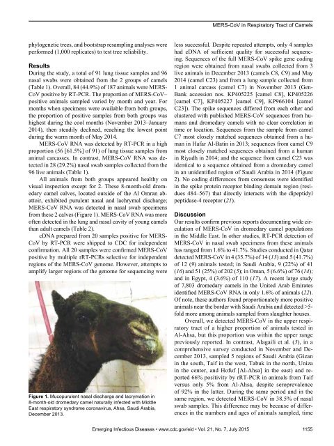
MERS-CoV in Respiratory Tract of Camelsphylogenetic trees, and bootstrap resampling analyses wereperformed (1,000 replicates) to test tree reliability.ResultsDuring the study, a total of 91 lung tissue samples and 96nasal swabs were obtained from the 2 groups of camels(Table 1). Overall, 84 (44.9%) of 187 animals were MERS-CoV positive by RT-PCR. The proportion of MERS-CoV–positive animals sampled varied by month and year. Formonths when specimens were available from both groups,the proportion of positive samples from both groups washighest during the cool months (November 2013–January2014), then steadily declined, reaching the lowest pointduring the warm month of May 2014.MERS-CoV RNA was detected by RT-PCR in a highproportion (56 [61.5%] of 91) of lung tissue samples fromanimal carcasses. In contrast, MERS-CoV RNA was detectedin 28 (29.2%) nasal swab samples collected from the96 live animals (Table 1).All animals from both groups appeared healthy onvisual inspection except for 2. These 8-month-old dromedarycamel calves, located outside of the Al Omran abattoir,exhibited purulent nasal and lachrymal discharge;MERS-CoV RNA was detected in nasal swab specimensfrom these 2 calves (Figure 1). MERS-CoV RNA was moreoften detected in the lung and nasal cavity of young camelsthan adult camels (Table 2).cDNA prepared from 20 samples positive for MERS-CoV by RT-PCR were shipped to CDC for independentconfirmation. All 20 samples were confirmed MERS-CoVpositive by multiple rRT-PCRs selective for independentregions of the MERS-CoV genome. However, attempts toamplify larger regions of the genome for sequencing wereFigure 1. Mucopurulent nasal discharge and lacrymation in8-month-old dromedary camel naturally infected with MiddleEast respiratory syndrome coronavirus, Ahsa, Saudi Arabia,December 2013.less successful. Despite repeated attempts, only 4 sampleshad cDNA of sufficient quality for successful sequencing.Sequences of the full MERS-CoV spike gene codingregion were obtained from nasal swabs collected from 3live animals in December 2013 (camels C8, C9) and May2014 (camel C23) and from a lung sample collected from1 animal carcass (camel C7) in November 2013 (Gen-Bank accession nos. KP405225 [camel C8], KP405226[camel C7], KP405227 [camel C9], KP966104 [camelC23]). The spike sequences differed from each other andclustered with published MERS-CoV sequences from humansand dromedary camels with no clear correlation intime or location. Sequences from the sample from camelC7 most closely matched sequences obtained from a humanin Hafar Al-Batin in 2013; sequences from camel C9most closely matched sequences obtained from a humanin Riyadh in 2014; and the sequence from camel C23 wasidentical to a sequence obtained from a dromedary camelin an unidentified region of Saudi Arabia in 2014 (Figure2). No coding differences from consensus were identifiedin the spike protein receptor binding domain region (residues484–567) that directly interacts with the dipeptidylpeptidase-4 receptor (21).DiscussionOur results confirm previous reports documenting wide circulationof MERS-CoV in dromedary camel populationsin the Middle East. In other studies, RT-PCR detection ofMERS-CoV in nasal swab specimens from these animalshas ranged from 1.6% to 41.7%. Studies conducted in Qatardetected MERS-CoV in 4 (35.7%) of 14 (13) and 5 (41.7%)of 12 (9) animals tested; in Saudi Arabia, 9 (22%) of 41(16) and 51 (25%) of 202 (5); in Oman, 5 (6.6%) of 76 (14);and in Egypt, 4 (3.6%) of 110 (17). A recent large studyof 7,803 dromedary camels in the United Arab Emiratesidentified MERS-CoV RNA in only 1.6% of animals (22).Of note, these authors found proportionately more positiveanimals near the border with Saudi Arabia and detected >5-fold more among animals sampled from slaughter houses.Overall, we detected MERS-CoV in the upper respiratorytract of a higher proportion of animals tested inAl-Ahsa, but this proportion was within the upper rangepreviously reported. In contrast, Alagaili et al. (5), in acomprehensive survey conducted in November and December2013, sampled 5 regions of Saudi Arabia (Gizanin the south, Taif in the west, Tabuk in the north, Unizain the center, and Hofuf [Al-Ahsa] in the east) and reported66% positivity by rRT-PCR in animals from Taifversus only 5% from Al-Ahsa, despite seroprevalenceof 92% in the latter. During the same period and in thesame region, we detected MERS-CoV in 38.5% of nasalswab samples. This difference may be because of differencesin the numbers and ages of animals sampled, timeEmerging Infectious Diseases • www.cdc.gov/eid • Vol. 21, No. 7, July 2015 1155
- Page 3 and 4:
July 2015SynopsisOn the CoverMarian
- Page 5 and 6:
1240 Gastroenteritis OutbreaksCause
- Page 7 and 8:
SYNOPSISDisseminated Infections wit
- Page 9 and 10: Disseminated Infections with Talaro
- Page 11 and 12: Disseminated Infections with Talaro
- Page 13 and 14: Macacine Herpesvirus 1 inLong-Taile
- Page 15 and 16: Macacine Herpesvirus 1 in Macaques,
- Page 17 and 18: Macacine Herpesvirus 1 in Macaques,
- Page 19: Macacine Herpesvirus 1 in Macaques,
- Page 23: Malaria among Young Infants, Africa
- Page 26 and 27: RESEARCHFigure 3. Dynamics of 19-kD
- Page 28 and 29: Transdermal Diagnosis of MalariaUsi
- Page 30 and 31: RESEARCHFigure 2. A) Acoustic trace
- Page 32 and 33: RESEARCHof malaria-infected mosquit
- Page 34 and 35: Lack of Transmission amongClose Con
- Page 36 and 37: RESEARCH(IFA) and microneutralizati
- Page 38 and 39: RESEARCHoropharyngeal, and serum sa
- Page 40 and 41: RESEARCH6. Assiri A, McGeer A, Perl
- Page 42 and 43: RESEARCHadvanced genomic sequencing
- Page 44 and 45: RESEARCHTable 2. Next-generation se
- Page 46 and 47: RESEARCHTable 3. Mutation analysis
- Page 48 and 49: RESEARCHReferences1. Baize S, Panne
- Page 50 and 51: Parechovirus Genotype 3 Outbreakamo
- Page 52 and 53: RESEARCHFigure 1. Venn diagramshowi
- Page 54 and 55: RESEARCHTable 2. HPeV testing of sp
- Page 56 and 57: RESEARCHFigure 5. Distribution of h
- Page 58 and 59: RESEARCHReferences1. Selvarangan R,
- Page 62 and 63: RESEARCHTable 2. Middle East respir
- Page 64 and 65: RESEARCHseroprevalence in domestic
- Page 66 and 67: RESEARCHmeasure their current surve
- Page 68 and 69: RESEARCHTable 2. States with labora
- Page 70 and 71: RESEARCHFigure 2. Comparison of sur
- Page 72 and 73: RESEARCH9. Centers for Disease Cont
- Page 74 and 75: RESEARCHthe analyses. Cases in pers
- Page 76 and 77: RESEARCHTable 3. Sampling results (
- Page 78 and 79: RESEARCHpresence of Legionella spp.
- Page 80 and 81: Seroprevalence for Hepatitis Eand O
- Page 82 and 83: RESEARCHTable 1. Description of stu
- Page 84 and 85: RESEARCHTable 3. Crude and adjusted
- Page 86 and 87: RESEARCHrates by gender or HIV stat
- Page 88 and 89: RESEARCH25. Taha TE, Kumwenda N, Ka
- Page 90 and 91: POLICY REVIEWDutch Consensus Guidel
- Page 92 and 93: POLICY REVIEWTable 3. Comparison of
- Page 94 and 95: POLICY REVIEW6. Botelho-Nevers E, F
- Page 96 and 97: DISPATCHESFigure 1. Phylogenetic tr
- Page 98 and 99: DISPATCHESSevere Pediatric Adenovir
- Page 100 and 101: DISPATCHESTable 1. Demographics and
- Page 102 and 103: DISPATCHES13. Kim YJ, Hong JY, Lee
- Page 104 and 105: DISPATCHESTable. Alignment of resid
- Page 106 and 107: DISPATCHESFigure 2. Interaction of
- Page 108 and 109: DISPATCHESSchmallenberg Virus Recur
- Page 110 and 111:
DISPATCHESFigure 2. Detection of Sc
- Page 112 and 113:
DISPATCHESFigure 1. Histopathologic
- Page 114:
DISPATCHESFigure 2. Detection of fo
- Page 117 and 118:
Influenza Virus Strains in the Amer
- Page 119 and 120:
Novel Arenavirus Isolates from Nama
- Page 121 and 122:
Novel Arenaviruses, Southern Africa
- Page 123 and 124:
Readability of Ebola Informationon
- Page 125 and 126:
Readability of Ebola Information on
- Page 127 and 128:
Patients under investigation for ME
- Page 129 and 130:
Patients under investigation for ME
- Page 131 and 132:
Wildlife Reservoir for Hepatitis E
- Page 133 and 134:
Asymptomatic Malaria and Other Infe
- Page 135 and 136:
Asymptomatic Malaria in Children fr
- Page 137 and 138:
Bufavirus in Wild Shrews and Nonhum
- Page 139 and 140:
Bufavirus in Wild Shrews and Nonhum
- Page 141 and 142:
Range Expansion for Rat Lungworm in
- Page 143 and 144:
Slow Clearance of Plasmodium falcip
- Page 145 and 146:
Slow Clearance of Plasmodium falcip
- Page 147 and 148:
Gastroenteritis Caused by Norovirus
- Page 149 and 150:
Ebola Virus Stability on Surfaces a
- Page 151 and 152:
Ebola Virus Stability on Surfaces a
- Page 153 and 154:
Outbreak of Ciprofloxacin-Resistant
- Page 155 and 156:
Outbreak of S. sonnei, South KoreaT
- Page 157 and 158:
Rapidly Expanding Range of Highly P
- Page 159 and 160:
Cluster of Ebola Virus Disease, Bon
- Page 161 and 162:
Cluster of Ebola Virus Disease, Lib
- Page 163 and 164:
ANOTHER DIMENSIONThe Past Is Never
- Page 165 and 166:
Measles Epidemic, Boston, Massachus
- Page 167 and 168:
LETTERSInfluenza A(H5N6)Virus Reass
- Page 169 and 170:
LETTERSsystem (8 kb-span paired-end
- Page 171 and 172:
LETTERS3. Van Hong N, Amambua-Ngwa
- Page 173 and 174:
LETTERSTable. Prevalence of Bartone
- Page 175 and 176:
LETTERSavian influenza A(H5N1) viru
- Page 177 and 178:
LETTERSprovinces and a total of 200
- Page 179 and 180:
LETTERS7. Manian FA. Bloodstream in
- Page 181 and 182:
LETTERSforward projections. N Engl
- Page 183 and 184:
LETTERS3. Guindon S, Gascuel OA. Si
- Page 185 and 186:
BOOKS AND MEDIAin the port cities o
- Page 187 and 188:
ABOUT THE COVERNorth was not intere
- Page 189 and 190:
Earning CME CreditTo obtain credit,
- Page 191:
Emerging Infectious Diseases is a p