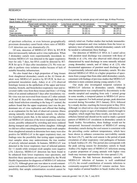
RESEARCHTable 2. Middle East respiratory syndrome coronavirus among dromedary camels, by sample group and camel age, Al-Ahsa Province,Saudi Arabia, 2013–2014*Nasal swab samples, live camels Lung tissue samples, camel carcasses Total samplesAge, yNo. tested No. (%) positive No. tested No. (%) positive No. tested No. (%) positiveYoung, 4 60 13 (21.7) 63 33 (52.4) 123 46 (37.4)Total samples 96 28 (29.2) 91 56 (61.5) 187 84 (44.9)*Tested by reverse transcription PCR.of specimen collection, or even between geographicallyproximate dromedary camel herds where rates of MERS-CoV detection can vary dramatically (9).Of note, detection of MERS-CoV RNA by RT-PCRdoes not necessarily indicate active virus replication. When3 dromedary camels were experimentally inoculated, infectiousMERS-CoV was detected in the upper respiratorytract for only 7 days, but RNA could be detected by RT-PCR for up to 35 days after inoculation (23). We were unableto perform virus isolation studies because of lack ofsuitable biosafety infrastructure.We also found that a high proportion of lung tissuesfrom slaughtered dromedary camels at the Al Omran abattoirwere MERS-CoV positive by RT-PCR. In their experimentalinoculation study, Adney et al. (23) observedhistologic lesions in the epithelium of the upper and lower(trachea, bronchi, and bronchioles) respiratory tract and recoveredviable virus from these tissues and from 1 of 4 lunglobes of an animal euthanized 5 days after inoculation; viablevirus was not recovered from tissues of 2 other animalsat 28 and 42 days after inoculation. Although that limitedstudy found infection extending to the lung of 1 animal, theauthors found that the upper respiratory tract was the predominantsite of virus replication and offered that findingas an explanation for the lack of observed systemic illnessamong naturally infected dromedary camels. An alternativehypothesis posits that, in the natural setting, subclinicalMERS-CoV infection of the lower respiratory tract alsooccurs, possibly enhanced by crowding and stress enduredduring transport and corralling before slaughter. Althoughwe did not collect matching premortem nasal swab samplesfrom slaughtered animals to determine how many were alsopositive for MERS-CoV in the upper respiratory tract, ourfindings raise the possibility that testing upper respiratorytract samples alone may underestimate the true numberof actively infected animals. In humans, MERS-CoV wasdetected in the lower respiratory tract of infected patientsfor ≈1 month while oronasal swab samples were negative(24). Likewise, MERS-CoV detection has been found tobe enhanced from lower respiratory tract specimens, andtherefore these specimens are recommended by the WorldHealth Organization for diagnosis of MERS-CoV infection(2,24,25). Although great care was taken to avoid contaminationwith ambient MERS-CoV present in the abattoir, thepossibility that sample contamination occurred cannot beentirely ruled out. Further studies that include immunohistologicexamination and virus isolation from the lower respiratorytract of naturally infected dromedary camels willbe needed to substantiate these findings.Our detection of MERS-CoV RNA in 2 camel calveswith purulent nasal discharge was consistent with those ofHemida et al. (16), who also observed mild clinical signscharacterized by nasal discharge in some naturally infectedyoung dromedary camels, and of Adney et al. (23), whodocumented appearance of purulent nasal discharge in the3 experimentally infected adult dromedary camels. We alsodetected MERS-CoV RNA in a higher proportion of specimensfrom younger than from older adult dromedary camels,consistent with findings of previous studies that MERS-CoVinfection is more common among young camels (5,16).Our study also investigated temporal variation inMERS-CoV infection in dromedary camels. Althoughdata interpretation was complicated by discontinuity in themonths sampled and sampling from only 1 animal groupin some months, a temporal pattern in MERS-CoV prevalencewas apparent. For both animal groups, peak detectionoccurred during November 2013–January 2014, followedby a steady decline, reaching the lowest point in May 2014.Although we observed no clear temporal differences in thegeographic origins or ages of dromedary camels brought toslaughter, which might bias these results, our data are neverthelesslimited and should not be used to imply a generalpattern of MERS-CoV circulation in dromedary camels inSaudi Arabia. Nevertheless, these findings would not beunexpected. Increased circulation of MERS-CoV amongdromedary camels during the cool season is consistent withthe prevailing cooler ambient temperatures, which havebeen shown to enhance coronavirus survivability outsidethe host (26,27), and the cool season is the period of peakcirculation of other respiratory viral pathogens of humansin Saudi Arabia (28–30). This period also corresponds withthe peak calving season for dromedary camels in SaudiArabia (16); higher rates of MERS-CoV infections amonga greater proportion of young animals with higher virusloads may increase opportunities for virus spread (5,16).Whereas the link between dromedary camels andMERS-CoV infection of humans is well established(15,31), the overall contribution of zoonotic infections tocommunity-acquired MERS-CoV remains unclear. Serologicstudies of animal handlers in Saudi Arabia who work1156 Emerging Infectious Diseases • www.cdc.gov/eid • Vol. 21, No. 7, July 2015
MERS-CoV in Respiratory Tract of Camelsin close proximity to dromedary camels have shown limitedevidence of MERS-CoV infection (32–34). Alghamdi etal. (35), who examined patterns of MERS-CoV infectionsamong humans in Saudi Arabia between June 2013 andMay 2014, did not find a concomitant temporal increasein human infections that corresponded with our findings indromedary camels. Those authors observed a slight, temporaryincrease in cases among humans in June and September2013 and few cases from October through February,after which cases and deaths sharply increased beginningin April 2014. The authors concluded that lower relativehumidity and higher temperatures during these monthsmight have contributed to the dramatic surge in reportedcases. However, more recent data from the World HealthOrganization (36) show a sharp decline in MERS-CoVcases among humans in May 2014; low numbers of caseswere reported from June through August 2014, when meantemperature was highest and relative humidity was lowestin Saudi Arabia (34). Moreover, a recent increase in numbersof MERS-CoV cases in humans from September 2014through February 2015 corresponds more closely with thetemporal pattern we found in dromedary camels the precedingyear. Further studies conducted over multiple yearsare needed to better understand the ecology of MERS-CoV,which might help inform intervention strategies to reducezoonotic infections.AcknowledgmentsWe thank Isam Al Jalii and Khalid Borsais for assistance withsample collection and Marzooq M. Al Eknah for financial support.Dr. Khalafalla is professor of veterinary virology at King FaisalUniversity, Al-Ahsa, Saudi Arabia. His research focus is on viraldiseases of dromedary camels.Figure 2. Midpoint-rooted phylogenetic tree of Middle Eastrespiratory syndrome coronavirus spike gene open reading framesequences of this virus obtained from camels and select humans(sequences available from GenBank). The estimated neighborjoiningtree was constructed from nucleotide alignments by usingMEGA <strong>version</strong> 6.06 (http://www.megasoftware.net). Sequencenames are derived from GenBank accession number | virus strainname | month-year of collection. Numbers in parentheses denotenumber of additional available identical spike gene sequencesobtained from same identified region of the representativestrains. Bootstrap support values (1,000 replicates) >70% areplotted at the indicated internal branch nodes. Scale bars indicatenumber of nucleotide substitutions per site. Sequences obtainedfrom camels are designated by an icon; sequences obtainedfrom camels in Al-Ahsa Province, Saudi Arabia, 2013–2014, aredesignated by an asterisk (*).References1. Zaki AM, van Boheemen S, Bestebroer TM, Osterhaus AD,Fouchier RA. Isolation of a novel coronavirus from a man withpneumonia in Saudi Arabia. N Engl J Med. 2012;367:1814–20.http://dx.doi.org/10.1056/NEJMoa12117212. Drosten C, Seilmaier M, Corman VM, Hartmann W, Scheible G,Sack S, et al. Clinical features and virological analysis of a case ofMiddle East respiratory syndrome coronavirus infection. LancetInfect Dis. 2013;13:745–51. http://dx.doi.org/10.1016/S1473-3099(13)70154-33. Memish ZA, Mishra N, Olival KJ, Fagbo SF, Kapoor V,Epstein JH, et al. Middle East respiratory syndrome coronavirus inbats, Saudi Arabia. Emerg Infect Dis. 2013;19:1819–23.http://dx.doi.org/10.3201/eid1911.1311724. Albarrak AM, Stephens GM, Hewson R, Memish ZA.Recovery from severe novel coronavirus infection. Saudi Med J.2012;33:1265–9.5. Alagaili AN, Briese T, Mishra N, Kapoor V, Sameroff SC, de Wit E,et al. Middle East respiratory syndrome coronavirus infection indromedary camels in Saudi Arabia. MBiol. 2014; e00884–14.6. Hemida MG, Perera RA, Wang P, Alhammadi MA, Siu LY, Li M,et al. Middle East respiratory syndrome (MERS) coronavirusEmerging Infectious Diseases • www.cdc.gov/eid • Vol. 21, No. 7, July 2015 1157
- Page 3 and 4:
July 2015SynopsisOn the CoverMarian
- Page 5 and 6:
1240 Gastroenteritis OutbreaksCause
- Page 7 and 8:
SYNOPSISDisseminated Infections wit
- Page 9 and 10:
Disseminated Infections with Talaro
- Page 11 and 12: Disseminated Infections with Talaro
- Page 13 and 14: Macacine Herpesvirus 1 inLong-Taile
- Page 15 and 16: Macacine Herpesvirus 1 in Macaques,
- Page 17 and 18: Macacine Herpesvirus 1 in Macaques,
- Page 19: Macacine Herpesvirus 1 in Macaques,
- Page 23: Malaria among Young Infants, Africa
- Page 26 and 27: RESEARCHFigure 3. Dynamics of 19-kD
- Page 28 and 29: Transdermal Diagnosis of MalariaUsi
- Page 30 and 31: RESEARCHFigure 2. A) Acoustic trace
- Page 32 and 33: RESEARCHof malaria-infected mosquit
- Page 34 and 35: Lack of Transmission amongClose Con
- Page 36 and 37: RESEARCH(IFA) and microneutralizati
- Page 38 and 39: RESEARCHoropharyngeal, and serum sa
- Page 40 and 41: RESEARCH6. Assiri A, McGeer A, Perl
- Page 42 and 43: RESEARCHadvanced genomic sequencing
- Page 44 and 45: RESEARCHTable 2. Next-generation se
- Page 46 and 47: RESEARCHTable 3. Mutation analysis
- Page 48 and 49: RESEARCHReferences1. Baize S, Panne
- Page 50 and 51: Parechovirus Genotype 3 Outbreakamo
- Page 52 and 53: RESEARCHFigure 1. Venn diagramshowi
- Page 54 and 55: RESEARCHTable 2. HPeV testing of sp
- Page 56 and 57: RESEARCHFigure 5. Distribution of h
- Page 58 and 59: RESEARCHReferences1. Selvarangan R,
- Page 60 and 61: RESEARCHthe left lobe was sampled b
- Page 64 and 65: RESEARCHseroprevalence in domestic
- Page 66 and 67: RESEARCHmeasure their current surve
- Page 68 and 69: RESEARCHTable 2. States with labora
- Page 70 and 71: RESEARCHFigure 2. Comparison of sur
- Page 72 and 73: RESEARCH9. Centers for Disease Cont
- Page 74 and 75: RESEARCHthe analyses. Cases in pers
- Page 76 and 77: RESEARCHTable 3. Sampling results (
- Page 78 and 79: RESEARCHpresence of Legionella spp.
- Page 80 and 81: Seroprevalence for Hepatitis Eand O
- Page 82 and 83: RESEARCHTable 1. Description of stu
- Page 84 and 85: RESEARCHTable 3. Crude and adjusted
- Page 86 and 87: RESEARCHrates by gender or HIV stat
- Page 88 and 89: RESEARCH25. Taha TE, Kumwenda N, Ka
- Page 90 and 91: POLICY REVIEWDutch Consensus Guidel
- Page 92 and 93: POLICY REVIEWTable 3. Comparison of
- Page 94 and 95: POLICY REVIEW6. Botelho-Nevers E, F
- Page 96 and 97: DISPATCHESFigure 1. Phylogenetic tr
- Page 98 and 99: DISPATCHESSevere Pediatric Adenovir
- Page 100 and 101: DISPATCHESTable 1. Demographics and
- Page 102 and 103: DISPATCHES13. Kim YJ, Hong JY, Lee
- Page 104 and 105: DISPATCHESTable. Alignment of resid
- Page 106 and 107: DISPATCHESFigure 2. Interaction of
- Page 108 and 109: DISPATCHESSchmallenberg Virus Recur
- Page 110 and 111: DISPATCHESFigure 2. Detection of Sc
- Page 112 and 113:
DISPATCHESFigure 1. Histopathologic
- Page 114:
DISPATCHESFigure 2. Detection of fo
- Page 117 and 118:
Influenza Virus Strains in the Amer
- Page 119 and 120:
Novel Arenavirus Isolates from Nama
- Page 121 and 122:
Novel Arenaviruses, Southern Africa
- Page 123 and 124:
Readability of Ebola Informationon
- Page 125 and 126:
Readability of Ebola Information on
- Page 127 and 128:
Patients under investigation for ME
- Page 129 and 130:
Patients under investigation for ME
- Page 131 and 132:
Wildlife Reservoir for Hepatitis E
- Page 133 and 134:
Asymptomatic Malaria and Other Infe
- Page 135 and 136:
Asymptomatic Malaria in Children fr
- Page 137 and 138:
Bufavirus in Wild Shrews and Nonhum
- Page 139 and 140:
Bufavirus in Wild Shrews and Nonhum
- Page 141 and 142:
Range Expansion for Rat Lungworm in
- Page 143 and 144:
Slow Clearance of Plasmodium falcip
- Page 145 and 146:
Slow Clearance of Plasmodium falcip
- Page 147 and 148:
Gastroenteritis Caused by Norovirus
- Page 149 and 150:
Ebola Virus Stability on Surfaces a
- Page 151 and 152:
Ebola Virus Stability on Surfaces a
- Page 153 and 154:
Outbreak of Ciprofloxacin-Resistant
- Page 155 and 156:
Outbreak of S. sonnei, South KoreaT
- Page 157 and 158:
Rapidly Expanding Range of Highly P
- Page 159 and 160:
Cluster of Ebola Virus Disease, Bon
- Page 161 and 162:
Cluster of Ebola Virus Disease, Lib
- Page 163 and 164:
ANOTHER DIMENSIONThe Past Is Never
- Page 165 and 166:
Measles Epidemic, Boston, Massachus
- Page 167 and 168:
LETTERSInfluenza A(H5N6)Virus Reass
- Page 169 and 170:
LETTERSsystem (8 kb-span paired-end
- Page 171 and 172:
LETTERS3. Van Hong N, Amambua-Ngwa
- Page 173 and 174:
LETTERSTable. Prevalence of Bartone
- Page 175 and 176:
LETTERSavian influenza A(H5N1) viru
- Page 177 and 178:
LETTERSprovinces and a total of 200
- Page 179 and 180:
LETTERS7. Manian FA. Bloodstream in
- Page 181 and 182:
LETTERSforward projections. N Engl
- Page 183 and 184:
LETTERS3. Guindon S, Gascuel OA. Si
- Page 185 and 186:
BOOKS AND MEDIAin the port cities o
- Page 187 and 188:
ABOUT THE COVERNorth was not intere
- Page 189 and 190:
Earning CME CreditTo obtain credit,
- Page 191:
Emerging Infectious Diseases is a p