th - 1988 - 51st ENC Conference
th - 1988 - 51st ENC Conference
th - 1988 - 51st ENC Conference
Create successful ePaper yourself
Turn your PDF publications into a flip-book with our unique Google optimized e-Paper software.
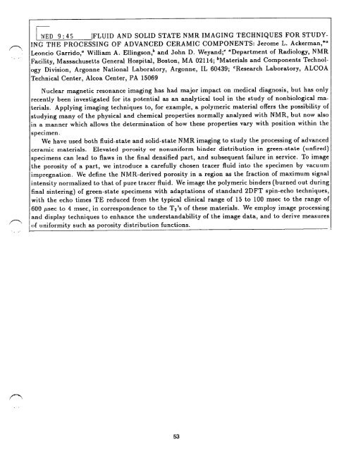
WED 9" 45 IFLUID AND SOLID STATE NMR IMAGING TECHNIQUES FOR STUDY-<br />
ING THE PROCESSING OF ADVANCED CERAMIC COMPONENTS: Jerome L. Ackerman, *~<br />
Leoncio Garrido, ~ William A. Ellingson, b and John D. Weyand; ~ ~Department of Radiology, NMR<br />
Facility, Massachusetts General Hospital, Boston, MA 02114; bMaterials and Components Technol-<br />
ogy Division, Argonne National Laboratory, Argonne, IL 60439; CResearch Laboratory, ALCOA<br />
Technical Center, Alcoa Center, PA 15069<br />
Nuclear magnetic resonance imaging has had major impact on medical diagnosis, but has only<br />
recently been investigated for its potential as an analytical tool in <strong>th</strong>e study of nonbiological ma-<br />
terials. Applying imaging techniques to, for example, a polymeric material offers <strong>th</strong>e possibility of<br />
studying many of <strong>th</strong>e physical and chemical properties normally analyzed wi<strong>th</strong> NMR, but now also<br />
in a manner which allows <strong>th</strong>e determination of how <strong>th</strong>ese properties vary wi<strong>th</strong> position wi<strong>th</strong>in <strong>th</strong>e<br />
specimen.<br />
We have used bo<strong>th</strong> fluid-state and solid-state NMR imaging to study <strong>th</strong>e processing of advanced<br />
ceramic materials. Elevated porosity or nonuniform binder distribution in green-state (unfired)<br />
specimens can lead to flaws in <strong>th</strong>e final densified part, and subsequent failure in service. To image<br />
<strong>th</strong>e porosity of a part, we introduce a carefully chosen tracer fluid into <strong>th</strong>e specimen by vacuum<br />
impregnation. We define <strong>th</strong>e NMR-derived porosity in a region as <strong>th</strong>e fraction of maximum signal<br />
intensity normalized to <strong>th</strong>at of pure tracer fluid. We image <strong>th</strong>e polymeric binders (burned out during<br />
final sintering) of green-state specimens wi<strong>th</strong> adaptations of standard 2DFT spin-echo techniques,<br />
wi<strong>th</strong> <strong>th</strong>e echo times TE reduced from <strong>th</strong>e typical clinical range of 15 to 100 msec to <strong>th</strong>e range of<br />
600 itsec to 4 msec, in correspondence to <strong>th</strong>e T2's of <strong>th</strong>ese materials. We employ image processing<br />
'and display techniques to enhance <strong>th</strong>e understandability of <strong>th</strong>e image data, and to derive measures<br />
of t, niformity such as porosity distribution functions.<br />
53


