

a la physique de l'information - Lisa - Université d'Angers
a la physique de l'information - Lisa - Université d'Angers
a la physique de l'information - Lisa - Université d'Angers

You also want an ePaper? Increase the reach of your titles
YUMPU automatically turns print PDFs into web optimized ePapers that Google loves.
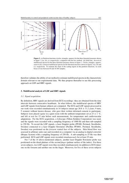
Multifractality in central and peripheral cardiovascu<strong>la</strong>r data of healthy subjects 621<br />
(a) (b)<br />
Figure 2. (a) Partition functions (circles, triangles, squares) for the three binomial measures shown<br />
in figure 1 ((a), (b), (c) respectively), computed with the box method. (b) Solid line: theoretical<br />
multifractal spectra for the three binomial measures shown in figure 1. Circles, triangles, squares:<br />
numerically estimated multifractal spectra for the binomial measures shown in figures 1(a), (b) and<br />
(c), respectively. To estimate the slope of the scaling region on the partition functions, we used<br />
boxes of size between 64 and 16 384 samples.<br />
therefore validates the ability of our method to estimate multifractal spectra in the characteristic<br />
domain relevant to our experimental data. We thus propose thereafter to use this processing<br />
approach on LDF and HRV signals.<br />
3. Multifractal analysis of LDF and HRV signals<br />
3.1. Signal acquisition<br />
By <strong>de</strong>finition, HRV signals are <strong>de</strong>rived from ECG recordings: they are obtained from the time<br />
intervals between consecutive heartbeats. In what follows, the multifractal spectra of HRV<br />
and LDF signals from human subjects are computed. The ECG and LDF signals processed in<br />
our work were recor<strong>de</strong>d simultaneously in 14 subjects (mean age 28.8 ± 11.2 years; 9 men,<br />
5 women) without known disease, who gave their written informed consent to participate.<br />
Subjects were p<strong>la</strong>ced supine in a quiet room with the ambient temperature set at 23 ± 1 ◦ C,<br />
and left at rest for 15 min before each measurement, for temperature and cardiovascu<strong>la</strong>r<br />
adaptations. For the ECG acquisition, a Lifescope (Nihon Koh<strong>de</strong>n Corporation) was used,<br />
and the signals were recor<strong>de</strong>d with a sampling frequency of 1000 Hz and then sub-sampled<br />
to 250 Hz. To record the LDF signals, a <strong>la</strong>ser Doppler probe (PF408, Perimed, Stockholm,<br />
Swe<strong>de</strong>n) connected to a <strong>la</strong>ser Doppler flowmeter (Periflux PF4001, Perimed, Stockholm,<br />
Swe<strong>de</strong>n) was positioned on the forearm ventral face of the subjects. Skin blood flow was<br />
assessed in arbitrary units (au) and recor<strong>de</strong>d on a computer via an analog-to-digital converter<br />
(Biopac System) with a sampling frequency of 250 Hz. A sub-sampling to 25 Hz was then<br />
performed. ECG and LDF signals were recor<strong>de</strong>d simultaneously foratleast25min.<br />
For 7 of the 14 above-mentioned subjects, another <strong>la</strong>ser Doppler flowmeter probe was<br />
positioned on the distal pha<strong>la</strong>nx of the second finger (palmar surface of the in<strong>de</strong>x). For these<br />
seven subjects, two LDF signals were thus recor<strong>de</strong>d simultaneously (in addition to ECG data):<br />
one on the forearm and another one on the finger. Moreover, for five of these seven subjects<br />
189/197