Protein Expression and Purification Series - Bio-Rad
Protein Expression and Purification Series - Bio-Rad
Protein Expression and Purification Series - Bio-Rad

Create successful ePaper yourself
Turn your PDF publications into a flip-book with our unique Google optimized e-Paper software.
<strong>Protein</strong> <strong>Expression</strong> <strong>and</strong> <strong>Purification</strong> <strong>Series</strong><br />
4.<br />
5.<br />
6.<br />
7.<br />
8.<br />
9.<br />
Run the gel at 200 V for 30 minutes. (If using a <strong>Bio</strong>-<strong>Rad</strong> Mini-PROTEAN TGX precast gel, the gel can<br />
be run at 300 V for 15 minutes.)<br />
After the run is complete, remove the gel from the cassette <strong>and</strong> place it in the gel staining tray.<br />
Wash the gel three times with 50 ml of distilled water for five minutes each rinse. Discard all the rinse<br />
water.<br />
Note: Make sure that all of the wash water has been removed since excess water diluting the gel stain<br />
will interfere with staining efficiency.<br />
Stain the gel with 50 ml of <strong>Bio</strong>-Safe Coomassie stain for one hour.<br />
After one hour discard the <strong>Bio</strong>-Safe Coomassie stain <strong>and</strong> add 100 ml of distilled water <strong>and</strong> destain the<br />
gel overnight.<br />
Image the gel if you have an imaging system, such as the <strong>Bio</strong>-<strong>Rad</strong> Gel Doc EZ system, or dry the<br />
gel if you have a cellophane drying system.<br />
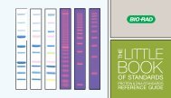
250 kD<br />
150<br />
100<br />
75<br />
50<br />
37<br />
25<br />
20<br />
15<br />
10<br />
Figure 4.2. Centrifugation purification process results analyzed by SDS-PAGE.<br />
Lane 1. Precision Plus Dual Color st<strong>and</strong>ards.<br />
Lane 2. Uninduced cells. There is no strong b<strong>and</strong> at approximately 43 kDa so no GST-DHFR-His has been expressed<br />
Lane 3. Induced cells. A strong b<strong>and</strong> is present at approximately 43 kDa that was not present in the uninduced cell sample (lane 2)<br />
<strong>and</strong> is GST-DHFR-His<br />
Lane 4. Insoluble fraction of E. coli cell lysate. A b<strong>and</strong> is present that is the same size as the induced b<strong>and</strong> in lane 3 <strong>and</strong> this<br />
represents the GST-DHFR-His that aggregated or was not folded properly <strong>and</strong> hence is not soluble <strong>and</strong> spins down with the cellular<br />
debris including cell walls, cell membranes, <strong>and</strong> the E. coli proteins which are not soluble in the 20 mM sodium phosphate, 300 mM<br />
NaCl, with 5 mM imidazole buffer<br />
Lane 5. Soluble fraction of E. coli cell lysate. A b<strong>and</strong> is present that is the same size as the induced b<strong>and</strong> in lane 3 <strong>and</strong> this<br />
represents the GST-DHFR-His which is soluble. The large b<strong>and</strong> at approximately 12 kDa is the lysozyme, which is also soluble, that<br />
was used to lyse the cells open.<br />
Lane 6. Flowthrough fraction which did not bind to the Ni-IMAC resin. This is the fraction of proteins from the soluble fraction<br />
that did not bind to the Ni-IMAC resin. The soluble fraction was suspended in 20 mM sodium phosphate buffer, 300 mM NaCl <strong>and</strong> 5<br />
mM imidazole. The high salt helped prevent E. coli proteins <strong>and</strong> the lysozyme from non-specifically sticking to the Ni-IMAC beads.<br />
The 5 mM imidazole helped prevent E. coli proteins with multiple histidine groups from binding to the Ni-IMAC beads. There is a<br />
decrease in the amount of GST-DHFR-His in the flowthrough fraction versus the soluble fraction (lane 5) <strong>and</strong> this is representative of<br />
the GST-DHFR-His binding to the Ni-IMAC resin.<br />
Lane 7. Wash fraction. This fraction contains proteins that were washed off the Ni-IMAC beads when a wash buffer that has a<br />
slightly higher imidazole level (10 mM) was added to wash off more non-specifically bound proteins. No GST-DHFR-His should wash<br />
off in this fraction since 10 mM imidazole is not enough to compete with the 6 histidine tag of GST-DHFR-His bound to the Ni-IMAC<br />
resin.<br />
Lane 8. Eluate fraction of GST-DHFR-His. This fraction contains the GST-DHFR-His. The elution buffer has 250 mM imidazole<br />
in it <strong>and</strong> this level of imidazole competes with the six histines of GST-DHFR-His <strong>and</strong> knocks them off the Ni sites <strong>and</strong> hence the<br />
GST-DHFR-His elutes or comes off the resin to be collected. This fraction is predominantly GST-DHFR-His protein relative to the<br />
unpurified soluble fraction in lane 5 that contains many other proteins.<br />
Lane 9. Desalted GST-DHFR-His. This fraction contains the purified GST-DHFR-His but has had the 250 mM imidazole removed.<br />
Also, since the desalting column removes smaller molecular weight compounds, some of the smaller molecular weight impurities<br />
found in lane 8 are not present in the desalted fraction.<br />
Chapter 4: 11 ml Culture Protocol for<br />
Centrifugation <strong>Purification</strong><br />
1 2 3 4 5 6 7 8 9<br />
107<br />
CHAPTER 4<br />
11 ml CULTURE<br />
PROTOCOL