Protein Expression and Purification Series - Bio-Rad
Protein Expression and Purification Series - Bio-Rad
Protein Expression and Purification Series - Bio-Rad

Create successful ePaper yourself
Turn your PDF publications into a flip-book with our unique Google optimized e-Paper software.
<strong>Protein</strong> <strong>Expression</strong> <strong>and</strong> <strong>Purification</strong> <strong>Series</strong><br />
weight. <strong>Protein</strong>s on the other h<strong>and</strong> are composed of 20 amino acids with molecular weights from 89 to<br />
204 Daltons (the average is 110). They vary considerably in amino acid composition. One Dalton equals<br />
the mass of a hydrogen atom, which is 1.66 x 10 -24 grams. Most proteins have masses on the order of<br />
thous<strong>and</strong>s of Daltons, so the term kilodalton (kD) is used for protein molecular masses. <strong>Protein</strong>s range in<br />
size from several kilodaltons to thous<strong>and</strong>s of kilodaltons but most fall between the range of 10 kD <strong>and</strong> 220<br />
kD. DHFR-GST-His has a primary structure of 410 amino acids, a total molecular weight of 52,000 daltons,<br />
or 52 kD.<br />
Using Gel Electrophoresis to Separate <strong>and</strong> Identify <strong>Protein</strong>s<br />
A protein’s electrical charge <strong>and</strong> its mass affect its mobility through a gel during electrophoresis. The ratio<br />
of charge to mass is called charge density. Since every protein is made of a unique combination of amino<br />
acids, the net charge of each protein may be different. The electric charge of proteins must be removed<br />
as a factor affecting migration in order for polyacrylamide electrophoresis to be effective as a method of<br />
protein molecular weight determination. The intrinsic charges of proteins are masked by placing a strongly<br />
anionic (negatively charged) detergent, sodium dodecyl sulfate<br />
(SDS), in both the sample buffer <strong>and</strong> the gel running buffer.<br />
SDS binds to <strong>and</strong> coats the proteins <strong>and</strong> also keeps them<br />
denatured as relatively linear chains. In this form, proteins<br />
migrate in a polyacrylamide gel as if they have equivalent<br />
negative charge densities, <strong>and</strong> mass becomes the main<br />
variable affecting the migration rate of each protein. (Note:<br />
Posttranslational modifications such as glycosylation can also<br />
affect protein migration).<br />
Chapter 1: Recombinant <strong>Protein</strong> <strong>Expression</strong> & <strong>Purification</strong><br />
47<br />
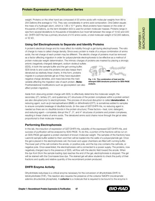
Fig. 1.13. The combination of heat <strong>and</strong> the<br />
detergent SDS denatures proteins for SDS-PAGE<br />
analysis.<br />
Aside from obscuring protein charge with SDS, to effectively determine the molecular weight, the<br />
secondary (2°), tertiary (3°), <strong>and</strong> quaternary (4°) structures of the protein complexes within a protein extract<br />
are also disrupted prior to electrophoresis. This process of structural disruption is called denaturation. A<br />
reducing agent, such as β-mercaptoethanol (BME) or dithiothreitol (DTT), is sometimes added to samples<br />
to ensure complete breakage of disulfide bonds. (In the case of GST-DHFR-His, no reducing agent is<br />
needed as there are no disulfide bonds in the protein structure.) Three factors—heat, ionic detergent,<br />
<strong>and</strong> reducing agent—completely disrupt the 2°, 3°, <strong>and</strong> 4° structures of proteins <strong>and</strong> protein complexes,<br />
resulting in linear chains of amino acids. The denatured amino acid chains move through the gel at rates<br />
proportional to their molecular masses.<br />
Performing Electrophoresis<br />
In this lab, the induction of expression of GST-DHFR-His, solubility of the expressed GST-DHFR-His, <strong>and</strong><br />
success of purification will be analyzed by SDS-PAGE. To do this, a portion of the fractions will be run on<br />
an SDS-PAGE gel against a protein st<strong>and</strong>ard of known molecular weight. The samples of the fractions have<br />
Laemmli sample buffer added to them <strong>and</strong> then will be loaded into the wells of a polyacrylamide gel. Once<br />
the gel is placed in the electrophoresis cell, the lower <strong>and</strong> upper chambers are filled with running buffer.<br />
The lower part of the cell contains the anode, or positive pole, <strong>and</strong> the top one contains the cathode, or<br />
negative pole. Once assembled, the electrophoresis cell is connected to a power supply. The proteins, now<br />
negatively charged due to the presence of SDS, will flow with the electric field toward the anode. When<br />
the dye front (from the sample loading dye) reaches the end of the gel, electrophoresis is stopped. The gel<br />
will be stained with the Coomassie blue dye. The stained gel will allow students to check the purity of their<br />
fractions <strong>and</strong> quality <strong>and</strong> relative quantity of the recombinant protein produced.<br />
DHFR Enzyme Activity<br />
Dihydrofolate reductase is a critical enzyme necessary for the conversion of dihydrofolate (DHF) to<br />
tetrahydrofolate (THF). This reaction also requires the presence of the cofactor NADPH (nicotinamide<br />
adenine dinucleotide phosphate). A cofactor is a molecule that is required to be bound to the enzyme in<br />
CHAPTER 1<br />
BACKGROUND