research activities in 2007 - CSEM
research activities in 2007 - CSEM
research activities in 2007 - CSEM

You also want an ePaper? Increase the reach of your titles
YUMPU automatically turns print PDFs into web optimized ePapers that Google loves.
Microfabricated Membranes for Cell Layer Culture and Analysis<br />
T. Overstolz, A. Hoogerwerf, M. Liley, F. Spano<br />
A microfabricated membrane chip is be<strong>in</strong>g developed for cell culture and analysis. Integrated Pt-electrodes allow the detection of <strong>in</strong>tercellular<br />
junctions and the evaluation of the ‘tightness” of epithelia cell layers. These tools are designed to screen the toxicity of nanoparticles, <strong>in</strong> particular<br />
their capacity to cross biological barriers such as the lungs.<br />
Nanomaterials based on nanoparticles have become very<br />
popular for many applications, <strong>in</strong>clud<strong>in</strong>g extremely strong<br />
composite materials based on carbon nanotubes. In parallel<br />
with this development, there has been a grow<strong>in</strong>g <strong>in</strong>terest to<br />
study the toxicity of these nanoparticles to the human body. In<br />
vitro methods for these tests are be<strong>in</strong>g developed <strong>in</strong> order to<br />
complement and/or replace large scale animal screen<strong>in</strong>g<br />
tests.<br />
In the body, epithelial cells are organized <strong>in</strong> sheets of cells<br />
that make up the epithelia. All epithelia have the function of<br />
provid<strong>in</strong>g a barrier between the body and the external world.<br />
In order to achieve this, <strong>in</strong>dividual epithelial cells are jo<strong>in</strong>ed via<br />
<strong>in</strong>tercellular junctions that make the epithelium impermeable.<br />
Thus, transport across the epithelia occurs essentially through<br />
the epithelial cells (trans-cellular transport) rather than<br />
between the cells (para-cellular transport).<br />
In vitro models of epithelia must be tested for the presence of<br />
<strong>in</strong>tercellular junctions and the absence of gaps between cells,<br />
to ensure that transport across the <strong>in</strong> vitro model closely<br />
resembles that of the epithelium <strong>in</strong> vivo. One of the most<br />
widely used approaches to determ<strong>in</strong>e the ‘tightness’ of a layer<br />
of cells is to measure its electrical impedance. Electrical<br />
impedances of around 200 Ohm/cm2 may be considered<br />
representative of “good” model epithelia that may be used to<br />
study transport processes.<br />
In order to m<strong>in</strong>imize the <strong>in</strong>fluence of artifacts that <strong>in</strong>terfere with<br />
impedance measurements <strong>in</strong> aqueous media (e.g.<br />
electrolysis, electrochemical potentials, foul<strong>in</strong>g of the<br />
electrode surface), the impedance of cell layers is usually<br />
measured us<strong>in</strong>g low frequency alternat<strong>in</strong>g currents <strong>in</strong> a<br />
4-term<strong>in</strong>al sens<strong>in</strong>g approach. In the 4-term<strong>in</strong>al sens<strong>in</strong>g<br />
method, one pair of electrodes is used to <strong>in</strong>ject the current <strong>in</strong>to<br />
the system, while a second pair of electrodes measures the<br />
potential across the cell layer.<br />
Figure 1: Silicon chip with <strong>in</strong>tegrated plat<strong>in</strong>um electrodes for<br />
impedance measurement of <strong>in</strong>tercellular junctions. The central cell<br />
culture well with its yellow porous Si3N4 membrane is visible. The two<br />
other square holes act as fluidics ports.<br />
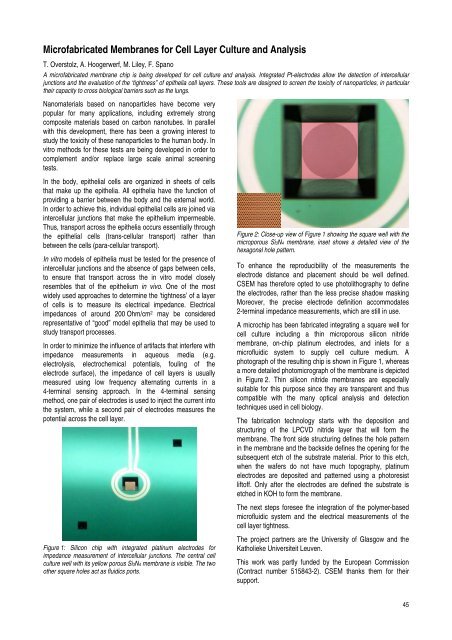
Figure 2: Close-up view of Figure 1 show<strong>in</strong>g the square well with the<br />
microporous Si3N4 membrane, <strong>in</strong>set shows a detailed view of the<br />
hexagonal hole pattern.<br />
To enhance the reproducibility of the measurements the<br />
electrode distance and placement should be well def<strong>in</strong>ed.<br />
<strong>CSEM</strong> has therefore opted to use photolithography to def<strong>in</strong>e<br />
the electrodes, rather than the less precise shadow mask<strong>in</strong>g<br />
Moreover, the precise electrode def<strong>in</strong>ition accommodates<br />
2-term<strong>in</strong>al impedance measurements, which are still <strong>in</strong> use.<br />
A microchip has been fabricated <strong>in</strong>tegrat<strong>in</strong>g a square well for<br />
cell culture <strong>in</strong>clud<strong>in</strong>g a th<strong>in</strong> microporous silicon nitride<br />
membrane, on-chip plat<strong>in</strong>um electrodes, and <strong>in</strong>lets for a<br />
microfluidic system to supply cell culture medium. A<br />
photograph of the result<strong>in</strong>g chip is shown <strong>in</strong> Figure 1, whereas<br />
a more detailed photomicrograph of the membrane is depicted<br />
<strong>in</strong> Figure 2. Th<strong>in</strong> silicon nitride membranes are especially<br />
suitable for this purpose s<strong>in</strong>ce they are transparent and thus<br />
compatible with the many optical analysis and detection<br />
techniques used <strong>in</strong> cell biology.<br />
The fabrication technology starts with the deposition and<br />
structur<strong>in</strong>g of the LPCVD nitride layer that will form the<br />
membrane. The front side structur<strong>in</strong>g def<strong>in</strong>es the hole pattern<br />
<strong>in</strong> the membrane and the backside def<strong>in</strong>es the open<strong>in</strong>g for the<br />
subsequent etch of the substrate material. Prior to this etch,<br />
when the wafers do not have much topography, plat<strong>in</strong>um<br />
electrodes are deposited and patterned us<strong>in</strong>g a photoresist<br />
liftoff. Only after the electrodes are def<strong>in</strong>ed the substrate is<br />
etched <strong>in</strong> KOH to form the membrane.<br />
The next steps foresee the <strong>in</strong>tegration of the polymer-based<br />
microfluidic system and the electrical measurements of the<br />
cell layer tightness.<br />
The project partners are the University of Glasgow and the<br />
Katholieke Universiteit Leuven.<br />
This work was partly funded by the European Commission<br />
(Contract number 515843-2). <strong>CSEM</strong> thanks them for their<br />
support.<br />
45