research activities in 2007 - CSEM
research activities in 2007 - CSEM
research activities in 2007 - CSEM

Create successful ePaper yourself
Turn your PDF publications into a flip-book with our unique Google optimized e-Paper software.
Detection Methods for Nanotoxicology<br />
S. Angeloni, V. Matera • , E. Verrecchia • , M. Liley<br />
An understand<strong>in</strong>g of the risks due to the short and long term toxicity of eng<strong>in</strong>eered nanoparticles requires the collection of a new body of data on<br />
nanoparticle toxicity. In vitro methods can contribute to the understand<strong>in</strong>g of the mechanisms by which NPs enter the human body, but require a<br />
sensitive nanoparticle detection technique. Inductively coupled plasma mass spectroscopy is a promis<strong>in</strong>g candidate technique.<br />
An assessment of the hazards due to eng<strong>in</strong>eered<br />
nanoparticles (NPs) is highly complex, requir<strong>in</strong>g extensive<br />
data collection on, among other th<strong>in</strong>gs, the toxicology of NPs.<br />
One approach that can contribute to this assessment is based<br />
on the use of absorption models deal<strong>in</strong>g with oral, <strong>in</strong>halation<br />
and topical absorption of NPs (via the <strong>in</strong>test<strong>in</strong>es, the lungs,<br />
and the sk<strong>in</strong>, respectively) [1] .<br />
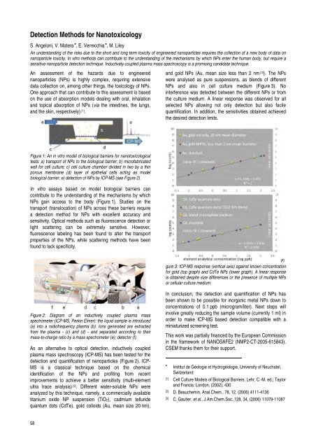
Figure 1: An <strong>in</strong> vitro model of biological barriers for nanotoxicological<br />
tests: a) transport of NPs to the biological barrier; b) microfabricated<br />
well for cell culture; c) cell culture chamber divided <strong>in</strong> two by a th<strong>in</strong><br />
porous membrane (d) layer of epithelial cells act<strong>in</strong>g as model<br />
biological barrier; e) detection of NPs by ICP-MS (see Figure 2).<br />
In vitro assays based on model biological barriers can<br />
contribute to the understand<strong>in</strong>g of the mechanisms by which<br />
NPs ga<strong>in</strong> access to the body (Figure 1). Studies on the<br />
transport (translocation) of NPs across these barriers require<br />
a detection method for NPs with excellent accuracy and<br />
sensitivity. Optical methods such as fluorescence detection or<br />
light scatter<strong>in</strong>g can be extremely sensitive. However,<br />
fluorescence label<strong>in</strong>g has been found to alter the transport<br />
properties of the NPs, while scatter<strong>in</strong>g methods have been<br />
found to lack specificity.<br />
Figure 2: Diagram of an <strong>in</strong>ductively coupled plasma mass<br />
spectrometer (ICP-MS, Perk<strong>in</strong> Elmer): the liquid sample is <strong>in</strong>troduced<br />
(a) <strong>in</strong>to a radiofrequency plasma (b). Ions generated are extracted<br />
from the plasma - (c) and (d) - and separated accord<strong>in</strong>g to their<br />
mass-to-charge ratio by a mass spectrometer (e); detector (f).<br />
As an alternative to optical detection, <strong>in</strong>ductively coupled<br />
plasma mass spectroscopy (ICP-MS) has been tested for the<br />
detection and quantification of nanoparticles (Figure 2). ICP-<br />
MS is a classical technique based on the chemical<br />
identification of the NPs and profit<strong>in</strong>g from recent<br />
improvements to achieve a better sensitivity (multi-element<br />
ultra trace analysis) [2] . Different water-soluble NPs were<br />
analyzed by this technique, namely, a commercially available<br />
titanium oxide NP suspension (TiO2), cadmium telluride<br />
quantum dots (CdTe), gold colloids (Au, mean size 20 nm),<br />
58<br />
and gold NPs (Au, mean size less than 2 nm [3] ). The NPs<br />
were analysed as pure suspensions, as blends of different<br />
NPs and also <strong>in</strong> cell culture medium (Figure 3). No<br />
<strong>in</strong>terference was detected between the different NPs or from<br />
the culture medium. A l<strong>in</strong>ear response was observed for all<br />
selected NPs allow<strong>in</strong>g not only detection but also facile<br />
quantification. In addition, the sensitivities obta<strong>in</strong>ed achieved<br />
the desired detection limits.<br />
Fi<br />
gure 3: ICP-MS response (vertical axis) aga<strong>in</strong>st known concentration<br />
for gold (top graph) and CdTe NPs (lower graph). A l<strong>in</strong>ear response<br />
is obta<strong>in</strong>ed despite size differences or the presence of multiple NPs<br />
or cellular culture medium.<br />
In conclusion, the detection and quantification of NPs has<br />
been shown to be possible for <strong>in</strong>organic metal NPs down to<br />
concentrations of 0.1 ppb (microgram/liter). Next steps will<br />
<strong>in</strong>volve greatly reduc<strong>in</strong>g the sample volume (currently 1 ml) <strong>in</strong><br />
order to make ICP-MS based detection compatible with a<br />
m<strong>in</strong>iaturized screen<strong>in</strong>g test.<br />
This work was partially f<strong>in</strong>anced by the European Commission<br />
<strong>in</strong> the framework of NANOSAFE2 (NMP2-CT-2005-615843).<br />
<strong>CSEM</strong> thanks them for their support.<br />
•<br />
Institut de Géologie et Hydrogéologie, University of Neuchatel,<br />
Switzerland<br />
[1] Cell Culture Models of Biological Barriers. Lehr, C.-M. ed.; Taylor<br />
and Francis: London, (2002), 430<br />
[2] D. Beauchem<strong>in</strong>, Anal.Chem., 78, 12, (2006) 4111-4136<br />
[3] C. Gautier, et al., J.Am.Chem.Soc.,128, 34, (2006) 11079-11087