research activities in 2007 - CSEM
research activities in 2007 - CSEM
research activities in 2007 - CSEM

Create successful ePaper yourself
Turn your PDF publications into a flip-book with our unique Google optimized e-Paper software.
Us<strong>in</strong>g Microtopography to Study Cell Elasticity<br />
M. Giazzon, N. Matthey, G. Weder, M. Tormen, T. Overstolz, M. Liley<br />
Tissue eng<strong>in</strong>eer<strong>in</strong>g and regenerative medic<strong>in</strong>e are of <strong>in</strong>creas<strong>in</strong>g importance as global life expectancy <strong>in</strong>creases. The development of surfaces to<br />
control cell adhesion and proliferation can make an important contribution to these fields. In this context, microtopographies are used <strong>in</strong> studies of<br />
the elasticity of human bone cells and as a tool to <strong>in</strong>fluence cell growth and survival.<br />
Micro and nano-topography can be used to promote or <strong>in</strong>hibit<br />
the proliferation of biological cells. One major field of<br />
application of this effect is that of tissue eng<strong>in</strong>eer<strong>in</strong>g and<br />
regenerative medic<strong>in</strong>e. The control of cell proliferation is of<br />
<strong>in</strong>creas<strong>in</strong>g importance <strong>in</strong> medical implants with the potential to<br />
accelerate their <strong>in</strong>tegration <strong>in</strong> the body, to reduce the risk of<br />
<strong>in</strong>fection or to avoid undesired cell growth <strong>in</strong> specific areas.<br />
For this reason a number of <strong>research</strong> groups are develop<strong>in</strong>g<br />
structured surfaces to improve and control the adhesion and<br />
proliferation of liv<strong>in</strong>g cells [1] .<br />
<strong>CSEM</strong> has been develop<strong>in</strong>g microtopographic structures <strong>in</strong><br />
order to study one specific aspect of cell behaviour that<br />
contributes to cell proliferation or death: cell elasticity. This is<br />
the capability of a cell to bend <strong>in</strong> response to a physical<br />
stimulus. Indeed cells may bend follow<strong>in</strong>g a stimulus or when<br />
they encounter a mechanical hurdle, but there is a limit above<br />
which they cannot curve [2] .Vary<strong>in</strong>g the microtopography<br />
allows that limit to be found.<br />
Microfabricated quartz surfaces with concentric grooves and<br />
ridges constitute a simple system for <strong>in</strong>vestigat<strong>in</strong>g cell<br />
elasticity. These substrates, obta<strong>in</strong>ed by photolithography,<br />
have ridges and grooves with a width of 2 or 4 µm and a<br />
range of depths from 30 nm to 500 nm. The radius of<br />
curvature of the ridges varies across the quartz substrate<br />
test<strong>in</strong>g cell elasticity under a range of conditions <strong>in</strong> one<br />
sample.<br />
When bone cells are grown on these surfaces they are found<br />
to assume different morphologies. Where the radius of<br />
curvature is high the cells adopt an elongated morphology<br />
(Figure 1b), follow<strong>in</strong>g the ridge and groove structure. In<br />
contrast, where the radius of curvature is small the cells<br />
spread over the grooves and ridges (Figure 1a).<br />
Figure 1: Bone cells on concentric grooves with a high curvature (a)<br />
and with a low curvature (b). Cells spread and orient normally on the<br />
flat surface <strong>in</strong> the top left of b. In the bottom right the cells are<br />
elongated and align to the grooves and ridges.<br />
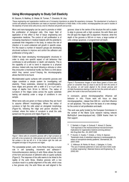
The cytoskeletal prote<strong>in</strong>, act<strong>in</strong>, forms fibres that play a crucial<br />
role <strong>in</strong> cell spread<strong>in</strong>g, movement and adhesion [3] .<br />
Immunofluorescence sta<strong>in</strong><strong>in</strong>g of act<strong>in</strong> fibres allows the study<br />
of the form and structure of adherent cells on quartz surfaces<br />
(Figure 2). The response of the cells seems to be l<strong>in</strong>ked to the<br />
rigidity of the act<strong>in</strong> fibres. Shallow grooves with a low<br />
curvature result <strong>in</strong> a partial alignment of the cells, which cross<br />
a few grooves only: the act<strong>in</strong> fibres are well aligned to the<br />
grooves: close to the centre of the structure the cell is unable<br />
to align to grooves with a high curvature: the act<strong>in</strong> fibers and<br />
the cell span the ridges with no alignment. However, when the<br />
depth of the grooves is 500 nm or more, a large number of<br />
cells undergo apoptosis, or programmed cell death.<br />
Figure 2: Fluorescence images of act<strong>in</strong> fibers (green) of bone cells<br />
grown on concentric l<strong>in</strong>es (a) close to and (b) far from the centre of<br />
the grooves. (c) cell nuclei aligned to the circular grooves and<br />
(d) triple fluorescence sta<strong>in</strong><strong>in</strong>g of cells far from the centre with act<strong>in</strong> <strong>in</strong><br />
green, the nuclei <strong>in</strong> blue and focal po<strong>in</strong>ts <strong>in</strong> red.<br />
In conclusion, groove microtopographies <strong>in</strong>fluence cell<br />
behavior <strong>in</strong> vitro. Future work will focus on deep<br />
microtopographies – deeper than 500 nm – and their <strong>in</strong>fluence<br />
on cell apoptosis. This may form the basis of a new strategy<br />
for the control of cell proliferation and death.<br />
This work was partly funded by the European Commission <strong>in</strong><br />
the context of the Marie Curie <strong>research</strong> tra<strong>in</strong><strong>in</strong>g network<br />
BioPolySurf (www.biopolysurf.net). <strong>CSEM</strong> thanks them for<br />
their support.<br />
[1] F. Rehfeldt, A. Engler, A. Eckhardt, F. Ahmed, D. Discher,<br />
”Cell responses to the mechanochemical microenvironment-<br />
Implications for regenerative medic<strong>in</strong>e and drug delivery”,<br />
Adv Drug Deliv Rev., 59, (<strong>2007</strong>)1329<br />
[2] P. Kunzler, C. Huwiler, T. Drobek, J. Vörös, N. Spencer,<br />
“Systematic study of osteoblast response to nanotopography by<br />
means of nanoparticle-density gradients”, Biomaterials, 28,<br />
(<strong>2007</strong>) 5000<br />
[3] C. Wilk<strong>in</strong>son, M. Riehle, M. Wood, J. Gallagher, A. Curtis,<br />
“The use of materials patterned on nano-and micro-metric scale<br />
<strong>in</strong> cellular eng<strong>in</strong>eer<strong>in</strong>g”, Materials Science and Eng<strong>in</strong>eer<strong>in</strong>g, 19,<br />
(2002) 263<br />
59