research activities in 2007 - CSEM
research activities in 2007 - CSEM
research activities in 2007 - CSEM

You also want an ePaper? Increase the reach of your titles
YUMPU automatically turns print PDFs into web optimized ePapers that Google loves.
Parallel Nanoscale Dispens<strong>in</strong>g of Liquids for Biological Analysis<br />
A. Meister, J. Przybylska, P. Niedermann, C. Santschi, M. Liley, H. He<strong>in</strong>zelmann<br />
Nanoscale dispens<strong>in</strong>g (NADIS) is a technique developed at <strong>CSEM</strong> to dispense ultrasmall volumes of liquid us<strong>in</strong>g specifically fabricated scann<strong>in</strong>g<br />
force microscopy probes. The NADIS probes consist of hollow cantilevers connected to a reservoir located <strong>in</strong> the chip body. Arrays of NADIS<br />
probes have been developed and produced by micromach<strong>in</strong><strong>in</strong>g <strong>in</strong> order to dispense biological liquids <strong>in</strong> parallel.<br />
The precise handl<strong>in</strong>g of liquids with volumes <strong>in</strong> the femto- and<br />
attoliter range is a challeng<strong>in</strong>g task, and one that is becom<strong>in</strong>g<br />
<strong>in</strong>creas<strong>in</strong>gly important for specific applications. For example,<br />
local surface functionalization with biomolecules is used to<br />
create high-density microarrays for diagnostics and biology. In<br />
the future, local application of candidate drugs or<br />
nanoparticles on biological cells may be used to study their<br />
<strong>in</strong>fluence on the cell <strong>in</strong> pharmacology or cytotoxicology.<br />
In <strong>CSEM</strong>’s nanoscale dispens<strong>in</strong>g, or “NADIS”, the ability to<br />
dispense ultrasmall volumes of liquid at precise positions on a<br />
sample is made possible by comb<strong>in</strong><strong>in</strong>g scann<strong>in</strong>g force<br />
microscopy (SFM) with a custom designed microtool. The<br />
NADIS microtool is similar to a standard SPM probe, except<br />
that the cantilever and the tip are hollow. The microfabrication<br />
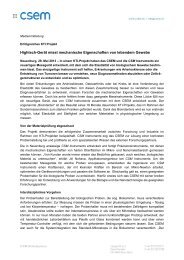
process for the NADIS cantilevers is shown <strong>in</strong> Figure 1.<br />
Figure 1: Fabrication process of the NADIS cantilever probes. a) A<br />
wafer is structured with reservoirs and V-grooves for chip release.<br />
b) A second wafer is processed for microfluidic channels and<br />
pyramidal tips with a silicon nitride layer. c) The pre-structured wafers<br />
are brought together and aligned. d) The wafers are fusion bonded<br />
and a silicon oxide layer is grown by thermal oxidation. e) The hollow<br />
cantilever is released by wet etch<strong>in</strong>g. f) The chip is released.<br />
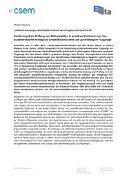
Figure 2: Example of glycerol droplets (left, optical micrograph)<br />
deposited with a tipless NADIS probe (right, SEM micrograph)<br />
The hollow core of the cantilever is <strong>in</strong>strumental for the<br />
dispens<strong>in</strong>g process as it connects an aperture <strong>in</strong> the cantilever<br />
tip to a liquid reservoir <strong>in</strong> the body of the chip. Once the<br />
reservoir is filled with liquid, the hollow cantilever and tip will<br />
be filled by capillarity. Transfer of liquid from the tip aperture to<br />
the surface occurs when the tip is brought <strong>in</strong>to contact with the<br />
sample. First proof of pr<strong>in</strong>ciple experiments demonstrat<strong>in</strong>g<br />
NADIS dispens<strong>in</strong>g with glycerol are shown <strong>in</strong> Figure 2.<br />
56<br />
In order to <strong>in</strong>crease NADIS throughput, systems with multiple<br />
cantilevers were developed. Various designs were def<strong>in</strong>ed,<br />
with different cantilever geometries and dimensions. Some of<br />
them are shown <strong>in</strong> Figure 3 and 4. Different microfluidic<br />
connections between the cantilevers and the reservoirs were<br />
also designed. All cantilevers <strong>in</strong> an array can be connected to<br />
one reservoir, so that all dispense the same liquid. Or, if<br />
different liquids are to be dispensed, each cantilever can be<br />
connected to its own reservoir. Cantilevers with a double<br />
beam structure are connected to an <strong>in</strong>let and an outlet<br />
reservoir, allow<strong>in</strong>g r<strong>in</strong>s<strong>in</strong>g of the cantilever by flush<strong>in</strong>g a liquid<br />
through it. Tests of these new systems are now underway.<br />
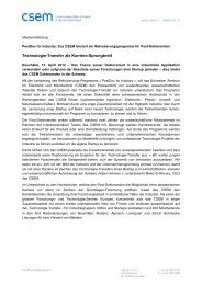
Figure 3: Optical micrograph of two chips with NADIS probe arrays of<br />
different designs (scale bar: 500 µm). The chips are still attached to<br />
the wafer.<br />
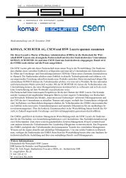
Figure 4: Scann<strong>in</strong>g electron microscope (SEM) micrographs of<br />
NADIS probes. a) Array of NADIS probes. b) Detail of an array.<br />
c) Close-up head-on view of a pyramidal shaped tip. d) Detail of a tip<br />
with the aperture at its apex.<br />
The partial support of the Swiss Federal Office for Education<br />
and Science (OFES) <strong>in</strong> the framework of the EC-funded<br />
Project NaPa (Contract no. NMP4-CT-2003-500120) is<br />
gratefully acknowledged.