research activities in 2007 - CSEM
research activities in 2007 - CSEM
research activities in 2007 - CSEM

You also want an ePaper? Increase the reach of your titles
YUMPU automatically turns print PDFs into web optimized ePapers that Google loves.
Sc<strong>in</strong>tillat<strong>in</strong>g Fiber Probes for Neurophysiology<br />
R. Eckert, B. Weber • , A. Buck • , M. Liley, R. Ischer, R. P. Stanley<br />
<strong>CSEM</strong> has been work<strong>in</strong>g with the University Hospital Zurich to develop optical probes for neurophysiology. The results of this development is a<br />
range of probes go<strong>in</strong>g from th<strong>in</strong> probes to be <strong>in</strong>serted <strong>in</strong>to tissue to large area probes to be used externally for long term measurements.<br />
Radioimag<strong>in</strong>g is widely used as a diagnostic tool <strong>in</strong> medic<strong>in</strong>e<br />
as it allows the direct imag<strong>in</strong>g of <strong>in</strong>ternal structures such as<br />
organs as well as their activity. Of these tools, positron<br />
emission tomography (PET) can give highly accurate images<br />
of the human body. PET has become extremely useful<br />
because compounds used <strong>in</strong> metabolism can be radiolabelled<br />
so that the signal strength is related to <strong>in</strong>ternal metabolic<br />
activity.<br />
Trac<strong>in</strong>g the evolution of these same compounds <strong>in</strong> time can<br />
also give an <strong>in</strong>sight <strong>in</strong>to the dynamics of metabolism. PET is<br />
too slow for this, a better option is to use a small probe that<br />
can take local measurements <strong>in</strong> real time. Together with the<br />
University Hospital Zurich and Swisstrace, <strong>CSEM</strong> has<br />
developed a range of fiber optic based probes for mak<strong>in</strong>g local<br />
physiological measurements.<br />
All fiber probes have two parts, a short part made from special<br />
fluorescent fibers and a standard glass or plastic optical fiber<br />
to transport the signal to the detection systems. The short<br />
fluorescent fibers convert ・ particles (electrons or positrons)<br />
com<strong>in</strong>g from radioactive decay that penetrate the fiber, <strong>in</strong>to<br />
light. The <strong>in</strong>tensity of the light depends on the energy of the<br />
beta particles. Only a very small quantity of light is produced –<br />
some tens of photons per beta particle.<br />
This light has to be guided without loss to a very low noise<br />
photodetection system. The fibre probes that have been<br />
developed have three different diameters, 250 µm, 500 µm<br />
and 3 mm. The th<strong>in</strong>nest fibers can be <strong>in</strong>serted <strong>in</strong>to tissue<br />
without caus<strong>in</strong>g damage, while the 3 mm probe has been<br />
designed as a surface probe for chronic (long term)<br />
measurements.<br />
The signal from the th<strong>in</strong>nest fiber is <strong>in</strong> the range of 30 counts<br />
per second (CPS) under standard operat<strong>in</strong>g conditions.<br />
However, ambient light can also enter the fiber, produc<strong>in</strong>g an<br />
unwanted background about ten orders of magnitude larger<br />
than the actual signal. In order to avoid work<strong>in</strong>g <strong>in</strong> complete<br />
darkness, the fibers have to be perfectly sealed aga<strong>in</strong>st stray<br />
light. Mak<strong>in</strong>g the fibers light tight is very challeng<strong>in</strong>g because<br />
the coat<strong>in</strong>g has to be completely opaque to light – even a<br />
s<strong>in</strong>gle micron-sized p<strong>in</strong>hole is unacceptable – while be<strong>in</strong>g th<strong>in</strong><br />
enough to allow the beta particles to reach the sc<strong>in</strong>tillat<strong>in</strong>g<br />
fiber.<br />
In order to achieve total light tightness, an opaque coat<strong>in</strong>g is<br />
applied <strong>in</strong> a layered fashion and is carefully tested after each<br />
layer to ensure specifications are achieved.<br />
The large 3 mm probes pose additional challenges. Due to<br />
their large area their cross section for ・ rays – which come<br />
from the dis<strong>in</strong>tegration of positrons (・ + particles) – is rather<br />
large. As the ・ rays can travel long distances, they are not a<br />
local signal and are therefore an unwanted noise. Optically it<br />
is impossible to tell the difference between fluorescence signal<br />
from the fiber com<strong>in</strong>g from ・‘s or ・’s. Given that the<br />
absorption length for ・ particles is much shorter than that of<br />
・ rays, the ratio between ・‘s and ・’s can be improved by<br />
th<strong>in</strong>n<strong>in</strong>g the fluorescent fiber to a th<strong>in</strong> disc 3 mm <strong>in</strong> diameter<br />
but less than 0.2 mm thick. These have been successfully<br />
used to monitor the differences <strong>in</strong> activity between the left and<br />
right hemispheres of the bra<strong>in</strong> <strong>in</strong> real time.<br />
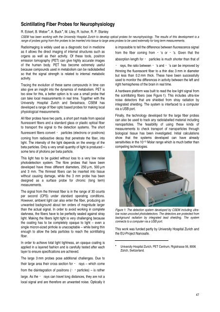
A hardware platform was built to read the low light signal from<br />
the sc<strong>in</strong>tillat<strong>in</strong>g fibers (see Figure 1). This <strong>in</strong>cludes ultra-low<br />
noise detectors that are shielded from stray radiation by<br />
<strong>in</strong>tegrated shield<strong>in</strong>g. The system is <strong>in</strong>terfaced to a computer<br />
via a USB port.<br />
F<strong>in</strong>ally, the technology developed for the large fiber probes<br />
can also be used to track any radiolabelled material <strong>in</strong>clud<strong>in</strong>g<br />
nanoparticles. The feasibility of us<strong>in</strong>g these k<strong>in</strong>ds of<br />
measurements to check transport of nanoparticles through<br />
biological tissue has been <strong>in</strong>vestigated. Initial calculations<br />
show that the systems developed can have already<br />
sensitivities <strong>in</strong> the 10-13 Molar range which is much better than<br />
compet<strong>in</strong>g technologies.<br />
Figure 1: The detection system developed by <strong>CSEM</strong> <strong>in</strong>clud<strong>in</strong>g ultralow<br />
noise uncooled photodetectors. The detectors are protected from<br />
background radiation by <strong>in</strong>tegrated lead shield<strong>in</strong>g. The system<br />
connects to a computer via a USB port.<br />
This work was funded partly by University Hospital Zurich and<br />
the EU-Project Nanosafe.<br />
•<br />
University Hospital Zurich, PET Centrum, Rigistrasse 56, 8006<br />
Zürich, Switzerland.<br />
47