

9%20ECOGRAFIA%20ABDOMINAL%20COMO%20CUANDO%20DONDE

You also want an ePaper? Increase the reach of your titles
YUMPU automatically turns print PDFs into web optimized ePapers that Google loves.
112<br />
ABDOMINAL ULTRASOUND<br />
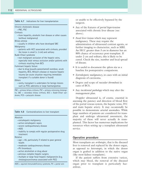
Table 4.7<br />
Indications for liver transplantation<br />
Chronic cholestatic disease<br />
—PBC, PSC<br />
Cirrhosis<br />
—from hepatitis, alcoholic liver disease or other causes<br />
(without malignancy)<br />
Biliary atresia<br />
—usually in children who have developed SBC<br />
Malignancy<br />
—patients with HCC associated with cirrhosis, provided<br />
the lesion is small (< 3 cm) and solitary<br />
Budd–Chiari syndrome<br />
—non-malignant occlusion of the hepatic veins,<br />
especially total venous occlusion and/or patients with<br />
cirrhosis resulting from BCS<br />
Fulminant hepatic failure<br />
—due to drug (usually paracetamol) overdose, acute<br />
hepatitis, BCS, Wilson’s disease or massive hepatic<br />
trauma (an acute situation requiring immediate<br />
transplant if a suitable donor is found)<br />
Others<br />
—rarely, transplant is undertaken for benign lesions<br />
such as PCD, adenoma or large haemangiomas<br />
PBC = primary biliary cirrhosis, PSC = primary sclerosing cholangitis,<br />
SBC = secondary biliary cirrhosis, BCS = Budd–Chiari syndrome,<br />
PCD = polycystic disease<br />
Table 4.8<br />
Contraindications to liver transplant<br />
Absolute<br />
—extrahepatic malignancy<br />
—active extrahepatic sepsis<br />
—severe cardiopulmonary disease<br />
—AIDS<br />
—inability to comply with regular postoperative drug<br />
treatment<br />
Relative<br />
—age > 65, particularly if related to poor general<br />
health<br />
—moderate cardiopulmonary disease<br />
—PV thrombosis<br />
—active alcoholism or drug abuse<br />
—previous complex hepatic surgery<br />
—multiple or large focal hepatic malignancies (e.g.<br />
cholangiocarcinomas associated with PSC)<br />
AIDS = aquired immunodeficiency syndrome, PV = portal vein<br />
or unable to be effectively bypassed by the<br />
surgeon.<br />
● Any of the features of portal hypertension<br />
associated with chronic liver disease (see<br />
above).<br />
● Focal liver lesions which may represent<br />
malignancy. These may require the<br />
administration of ultrasound contrast agents, or<br />
further imaging to characterize, such as MRI.<br />
An HCC greater than 3 cm in diameter has an<br />
80% chance of recurrence post-transplant. If<br />
under 2 cm and solitary, this is likely to be<br />
cured. Check the size, number and local spread<br />
of disease.<br />
● It is useful to document the spleen size as a<br />
baseline for postoperative comparisons.<br />
● Extrahepatic malignancy, in cases with an initial<br />
diagnosis of carcinoma.<br />
● Degree and scope of vascular thrombosis in<br />
cases of BCS.<br />
● Any incidental pathology which may alter the<br />
management plan.<br />
Doppler ultrasound is, of course, essential in<br />
assessing the patency and direction of blood flow<br />
of the portal venous system, the hepatic veins, IVC<br />
and main hepatic artery. It may occasionally be<br />
possible to demonstrate arterial anomalies. While<br />
large numbers of patients are considered for transplant<br />
and undergo ultrasound assessment, the<br />
majority of these will never actually be transplanted.<br />
This factor has numerous implications for<br />
resources when setting up a transplant ultrasound<br />
service.<br />
Operative procedure<br />
Most transplants are orthotopic, that is the diseased<br />
liver is removed and replaced by the donor organ,<br />
as opposed to heterotopic, in which the donor<br />
organ is grafted in addition to the native organ<br />
(like most kidney transplants).<br />
If the patient suffers from extensive varices,<br />
which may bleed, the removal of the diseased<br />
organ prior to transplant is particularly hazardous.