

9%20ECOGRAFIA%20ABDOMINAL%20COMO%20CUANDO%20DONDE

Create successful ePaper yourself
Turn your PDF publications into a flip-book with our unique Google optimized e-Paper software.
PATHOLOGY OF THE GALLBLADDER AND BILIARY TREE 67<br />
phosphatase and bilirubin levels should be normal in<br />
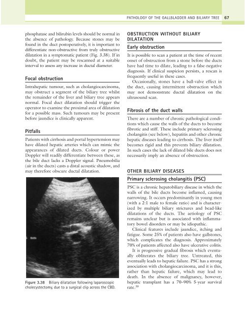
the absence of pathology. Because stones may be<br />
found in the duct postoperatively, it is important to<br />
differentiate non-obstructive from truly obstructive<br />
dilatation in a symptomatic patient (Fig. 3.38). If in<br />
doubt, the patient may be rescanned at a suitable<br />
interval to assess any increase in ductal diameter.<br />
Focal obstruction<br />
Intrahepatic tumour, such as cholangiocarcinoma,<br />
may obstruct a segment of the biliary tree whilst<br />
the remainder of the liver and biliary tree appears<br />
normal. Focal duct dilatation should trigger the<br />
operator to examine the proximal area of dilatation<br />
for a possible mass. Such tumours may be present<br />
before jaundice is clinically apparent.<br />
Pitfalls<br />
Patients with cirrhosis and portal hypertension may<br />
have dilated hepatic arteries which can mimic the<br />
appearances of dilated ducts. Colour or power<br />
Doppler will readily differentiate between these, as<br />
the bile duct lacks a Doppler signal. Pneumobilia<br />
(air in the ducts) casts a distal acoustic shadow, and<br />
may therefore obscure ductal dilatation.<br />
Figure 3.38 Biliary dilatation following laparoscopic<br />
cholecystectomy, due to a surgical clip across the CBD.<br />
OBSTRUCTION WITHOUT BILIARY<br />
DILATATION<br />
Early obstruction<br />
It is possible to scan a patient at the time of recent<br />
onset of obstruction from a stone before the ducts<br />
have had time to dilate, leading to a false-negative<br />
diagnosis. If clinical suspicion persists, a rescan is<br />
frequently useful in these cases.<br />
Occasionally, stones have a ball-valve effect in<br />
the duct, causing intermittent obstruction which<br />
may not demonstrate ductal dilatation on the<br />
ultrasound scan.<br />
Fibrosis of the duct walls<br />
There are a number of chronic pathological conditions<br />
which cause the walls of the ducts to become<br />
fibrotic and stiff. These include primary sclerosing<br />
cholangitis (see below), hepatitis and other chronic<br />
hepatic diseases leading to cirrhosis. The liver itself<br />
becomes rigid and this prevents biliary dilatation.<br />
In such cases the lack of dilated bile ducts does not<br />
necessarily imply an absence of obstruction.<br />
OTHER BILIARY DISEASES<br />
Primary sclerosing cholangitis (PSC)<br />
PSC is a chronic hepatobiliary disease in which the<br />
walls of the bile ducts become inflamed, causing<br />
narrowing. It occurs predominantly in young men<br />
(with a 2:1 male to female ratio) and is characterized<br />
by multiple biliary strictures and bead-like<br />
dilatations of the ducts. The aetiology of PSC<br />
remains unclear but is associated with inflammatory<br />
bowel disorders or may be idiopathic.<br />
Clinical features include jaundice, itching and<br />
fatigue. Some 25% of patients also have gallstones,<br />
which complicates the diagnosis. Approximately<br />
70% of patients affected also have ulcerative colitis.<br />
It is progressive gradual fibrosis which eventually<br />
obliterates the biliary tree. Untreated, this<br />
eventually leads to hepatic failure. PSC has a strong<br />
association with cholangiocarcinoma, and it is this,<br />
rather than hepatic failure, which may lead to<br />
death. In the absence of malignancy, however,<br />
hepatic transplant has a 70–90% 5-year survival<br />
rate. 30