

9%20ECOGRAFIA%20ABDOMINAL%20COMO%20CUANDO%20DONDE

You also want an ePaper? Increase the reach of your titles
YUMPU automatically turns print PDFs into web optimized ePapers that Google loves.
INTERVENTIONAL AND OTHER TECHNIQUES 255<br />
Methods of ultrasound guidance<br />
There are various ways of performing ultrasoundguided<br />
procedures: organ/lesion localization<br />
(‘blind biopsy’), biopsy guide or freehand technique.<br />
The choice of method depends upon the<br />
procedure in question, equipment and the experience<br />
and skill of the operator.<br />
Blind biopsy<br />
With this method a position on the skin surface is<br />
marked overlying the organ or lesion to be biopsied,<br />
using ultrasound to localize. This remains acceptable<br />
for diffuse disease, when only a representative<br />
sample of liver tissue is required. Nevertheless, it is<br />
good practice even in these situations to visualize<br />
the needle during the procedure, and this method<br />
of biopsy is now used less frequently.<br />
Biopsy guidance<br />
Most manufacturers provide a biopsy guide which<br />
fits snugly on to the transducer head and provides<br />
a rigid pathway for the needle (Fig. 11.1). These<br />
are now the commonest and preferred method of<br />
biopsy. Previously adjustable angle biopsy guides<br />
were available; however these offered no specific<br />
advantages and were prone to user error. The fixed<br />
biopsy guides contain a groove for a series of plastic<br />
inserts ranging from 14G to 22G size, depending<br />
on the size of the biopsy needle. It is often preferred<br />
to use one size greater than the needle, that<br />
is a 16G insert for an 18G needle, as the needle<br />
tends to move more freely. These guides are sterilized<br />
and fitted on to the transducer, which can<br />
either be covered by a sterile sheath or thoroughly<br />
cleaned with chlorhexidine solution. The use of a<br />
sheath is highly recommended, as it maintains the<br />
sterility of the procedure, reducing the risk of<br />
infection, with no adverse effect on the image.<br />
The needle pathway is displayed on the ultrasound<br />
monitor electronically as a line or narrow<br />
sector, through which the needle passes. The operator<br />
then scans in order to align the electronic<br />
pathway along the chosen route, the needle is<br />
inserted and the biopsy taken. These attachments<br />
should be tested regularly to ensure the needle follows<br />
the correct path (Fig. 11.2).<br />
Freehand<br />
A freehand approach, in which the operator scans<br />
with one hand and introduces the needle near to<br />
the transducer with the other, may be used for<br />
larger or more superficial lesions. This technique is<br />
commonly used for breast biopsy and biopsy in the<br />
head and neck. The needle is inserted from one<br />
end of the probe at right angles to the ultrasound<br />
beam; generally speaking the angle utilized is shallow<br />
in comparison with the fixed guide systems for<br />
deeper structures.<br />
d<br />
C<br />
a<br />
b<br />
A<br />
B<br />
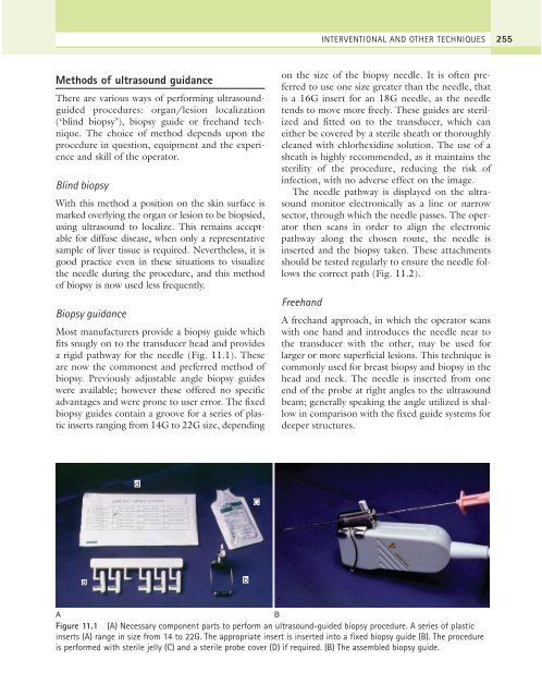
Figure 11.1 (A) Necessary component parts to perform an ultrasound-guided biopsy procedure. A series of plastic<br />
inserts (A) range in size from 14 to 22G. The appropriate insert is inserted into a fixed biopsy guide (B). The procedure<br />
is performed with sterile jelly (C) and a sterile probe cover (D) if required. (B) The assembled biopsy guide.