

9%20ECOGRAFIA%20ABDOMINAL%20COMO%20CUANDO%20DONDE

Create successful ePaper yourself
Turn your PDF publications into a flip-book with our unique Google optimized e-Paper software.
116<br />
ABDOMINAL ULTRASOUND<br />
Figure 4.32 The site of anastomosis in the IVC in a<br />
liver transplant.<br />
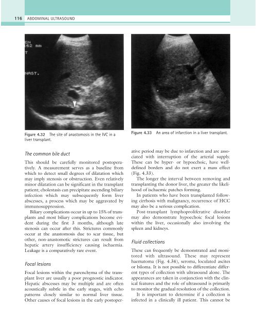
Figure 4.33<br />
An area of infarction in a liver transplant.<br />
The common bile duct<br />
This should be carefully monitored postoperatively.<br />
A measurement serves as a baseline from<br />
which to detect small degrees of dilatation which<br />
may imply stenosis or obstruction. Even relatively<br />
minor dilatation can be significant in the transplant<br />
patient; cholestasis can precipitate ascending biliary<br />
infection which may subsequently form liver<br />
abscesses, a process which may be aggravated by<br />
immunosuppression.<br />
Biliary complications occur in up to 15% of transplants<br />
and most biliary complications become evident<br />
during the first 3 months, although late<br />
stenosis can occur after this. Strictures commonly<br />
occur at the anastomosis due to scar tissue, but<br />
other, non-anastomotic strictures can result from<br />
hepatic artery insufficiency causing ischaemia.<br />
Leakage is a comparatively rare event.<br />
Focal lesions<br />
Focal lesions within the parenchyma of the transplant<br />
liver are usually a poor prognostic indicator.<br />
Hepatic abscesses may be multiple and are often<br />
acoustically subtle in the early stages, with echo<br />
patterns closely similar to normal liver tissue.<br />
Other causes of focal lesions in the early postoperative<br />
period may be due to infarction and are associated<br />
with interruption of the arterial supply.<br />
These can be hyper- or hypoechoic, have welldefined<br />
borders and do not exert a mass effect<br />
(Fig. 4.33).<br />
The longer the interval between removing and<br />
transplanting the donor liver, the greater the likelihood<br />
of ischaemic patches forming.<br />
In patients who have been transplanted following<br />
cirrhosis with malignancy, recurrence of HCC<br />
may also be a serious complication.<br />
Post-transplant lymphoproliferative disorder<br />
may also demonstrate hypoechoic focal lesions<br />
within the liver, occasionally also involving the<br />
spleen and kidneys.<br />
Fluid collections<br />
These can frequently be demonstrated and monitored<br />
with ultrasound. These may represent<br />
haematoma (Fig. 4.34), seroma, loculated ascites<br />
or biloma. It is not possible to differentiate different<br />
types of collection with ultrasound alone. The<br />
appearances are taken in conjunction with the clinical<br />
features and the role of ultrasound is primarily<br />
to monitor the gradual resolution of the collection.<br />
It is important to determine if a collection is<br />
infected in a clinically ill patient. This cannot be