

9%20ECOGRAFIA%20ABDOMINAL%20COMO%20CUANDO%20DONDE

You also want an ePaper? Increase the reach of your titles
YUMPU automatically turns print PDFs into web optimized ePapers that Google loves.
48<br />
ABDOMINAL ULTRASOUND<br />
ENLARGEMENT OF THE GALLBLADDER<br />
Because of the enormous variation in size and<br />
shape of the normal gallbladder, it is not possible<br />
to diagnose pathological enlargement by simply<br />
using measurements. Three-dimensional techniques<br />
may prove useful in assessing gallbladder<br />
volume 6 but this is a technique which is only likely<br />
to be clinically useful in a minority of patients with<br />
impaired gallbladder emptying.<br />
An enlarged gallbladder is frequently referred to<br />
as hydropic. It may be due to obstruction of the<br />
cystic duct (see below) or associated with numerous<br />
disease processes such as diabetes, primary<br />
sclerosing cholangitis, leptospirosis or in response<br />
to some types of drug.<br />
A pathologically dilated gallbladder, as opposed<br />
to one which is physiologically dilated, usually<br />
assumes a more rounded, tense appearance.<br />
B<br />
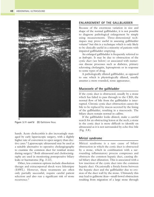
Figure 3.11 cont’d<br />
(B) Gallstone Ileus.<br />
hands. Acute cholecystitis is also increasingly managed<br />
by early laparoscopic surgery, with a slightly<br />
higher rate of conversion to open surgery than elective<br />
cases. 3 Laparoscopic ultrasound may be used as<br />
a suitable alternative to operative cholangiography<br />
to examine the common duct for residual stones<br />
during surgery. 4 Both ultrasound and cholescintigraphy<br />
are used in monitoring postoperative biliary<br />
leaks or haematoma (Fig. 3.13).<br />
Other, less common options include dissolution<br />
therapy and extracorporeal shock wave lithotripsy<br />
(ESWL). However, these treatments are often<br />
only partially successful, require careful patient<br />
selection and also run a significant risk of stone<br />
recurrence. 5<br />
Mucocoele of the gallbladder<br />
If the cystic duct is obstructed, usually by a stone<br />
which has failed to pass through to the CBD, the<br />
normal flow of bile from the gallbladder is interrupted.<br />
Chronic cystic duct obstruction causes the<br />
bile to be replaced by mucus secreted by the lining<br />
of the gallbladder, resulting in a mucocoele. The<br />
biliary ducts remain normal in calibre.<br />
If the gallbladder looks dilated, make a careful<br />
search for an obstructing lesion at the neck; a stone<br />
in the cystic duct is more difficult to identify on<br />
ultrasound as it is not surrounded by echo-free bile<br />
(Fig. 3.8).<br />
Mirizzi syndrome<br />
Mirizzi syndrome is a rare cause of biliary<br />
obstruction in which the cystic duct is obstructed<br />
by a stone, which in combination with a surrounding<br />
inflammatory process compresses and<br />
obstructs the common hepatic duct, causing distal<br />
biliary duct dilatation. This is associated with a<br />
low insertion of the cystic duct into the common<br />
hepatic duct. Occasionally a fistula forms between<br />
the hepatic duct and the gallbladder due to erosion<br />
of the duct wall by the stone. Ultimately this<br />
may lead to gallstone ileus—small-bowel obstruction<br />
resulting from migration of a large stone through